Microscope is an instrument that magnifies objects and details too small to be readily seen by the naked eye. The microscope ranks as one of the most important scientific tools. Physicians and biologists, for example, use microscopes to examine cells and tissues, and to look for evidence of infection within cells. Biology students learn about algae, protozoa, and other single-celled organisms by studying them under a microscope. Materials scientists and engineers use microscopes to study the crystal structures within metals. Modern computer chips have structures that are only visible with microscopes.
There are four basic kinds of microscopes: (1) optical, also called light; (2) electron; (3) scanning probe; and (4) ion. Much of this article discusses optical microscopes.
Optical microscopes
Optical microscopes work using lenses. A lens is a transparent object that refracts (bends) light. The simplest optical microscope is a magnifying glass. A magnifying glass has only one lens. The most powerful magnifying glasses can magnify an object by 10 to 20 times. A compound microscope uses two or more lens systems to provide higher magnifications. A lens system is a set of lenses that work together to magnify the image.
Magnification power is expressed using a number followed by the symbol X. For example, a 10X magnifying glass magnifies a specimen by 10 times. Resolution, or resolving power, is a measure that describes the ability to produce a clear image. Images produced by low- resolution microscopes tend to appear blurry, even if the images are highly magnified.
Basic optical microscopes.
Most optical microscopes used in teaching have three main parts. They are: (1) the tube, which contains the magnifying lenses; (2) the body, which supports the microscope; and (3) the stage, which holds the sample being viewed.
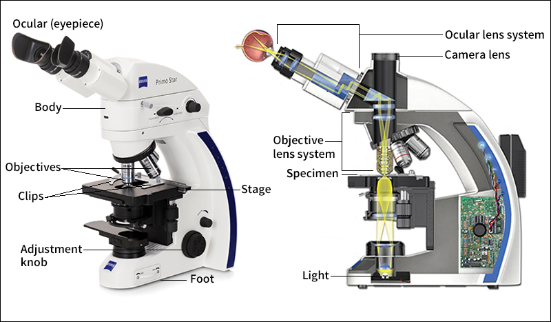
Tube and lenses.
A compound microscope’s tube contains two different kinds of lens systems. The lens system at the top, closest to the viewer’s eye, is called the ocular or eyepiece. At the opposite end of the tube, closest to the sample, is the objective. Microscopes often have several objective lens systems. These objectives are mounted in a rotating nosepiece. Each objective provides a different level of magnification. The viewer can rotate the nosepiece to choose the desired magnification level. For example, many simple compound microscopes have three objectives, with magnifications of 4X, 10X, and 40X. These objectives work with a 10X ocular. Thus, such a microscope can provide total magnifications of 40X, 100X, and 400X.
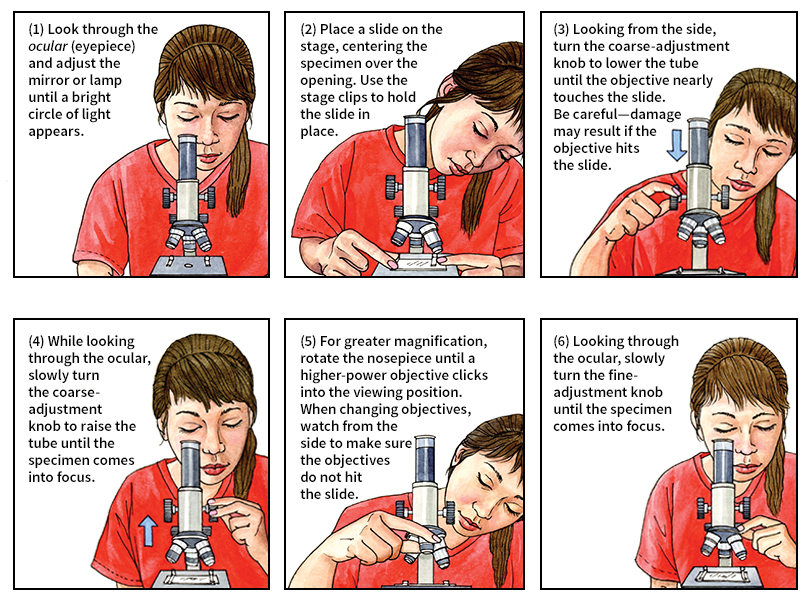
Optical microscopes used in research have lens systems that are much more powerful. Many such microscopes have a 100X objective and a 20X ocular, providing a total magnification of 2,000X. Nearly all high-powered research microscopes have oil immersion objectives. To use such an objective, the researcher places a drop of special oil on the slide, touching the bottom objective lens. The oil refracts light in a way that helps improve resolution at high magnification.
The body
consists of a base and an upright support that holds the tube. Knobs enable the viewer to move the lens tube closer to or farther from the sample. At the lower end of the body is a light source. Light from this source shines up through the sample. The light is refracted by the tube’s lenses and eventually enters the viewer’s eye.
The stage
is a platform that holds the sample above the light source. Samples are mounted on glass slides that measure 3 inches (76 millimeters) long and 1 inch (25 millimeters) wide. An opening in the stage allows light to illuminate the sample.
Many samples viewed through a microscope are transparent. However, samples are often stained with different dyes. The dyes may make it easier to see structures within cells. The technique of preparing samples for viewing with a microscope is called microtomy.
Specialized optical microscopes and techniques
enable scientists to view enhanced details. Specialized microscopes may manipulate the light used to illuminate the sample and use computers to gather and process the image.
Fluorescence microscopy,
a technique commonly used by biologists and medical researchers, usually involves staining samples with dyes that emit (give off) colored light. The light is typically captured by a digital camera or another detector connected to the microscope. By using dyes effectively and manipulating the captured images, scientists can identify tiny details within a cell.
Scanning optical microscopes
do not illuminate the entire sample at once. Instead, the microscope directs a laser beam at one small part of the sample at a time. A device called a photodetector measures the laser light shining through—or reflected from—the sample at each spot. After the beam passes over the whole sample, a computer combines the measurements, producing an image. If a sample’s depth is measured in this way, the result may be a three-dimensional image that can be viewed at multiple angles on a computer.
Phase contrast microscopes
make use of variations in the phase (alignment) of light waves. Such variations are created as light passes through different substances or through samples of varying thickness. A phase contrast microscope makes variations in phase appear as variations in brightness. In this way, the microscope can make visible parts of a transparent object that vary in thickness or that have certain other optical properties.
Dark field microscopes
prevent light from the lamp from shining directly up the tube. Instead, the microscope uses only light scattered by the specimen. The specimen appears bright against a black background.
Other kinds of microscopes
Because of the nature of light, optical microscopes lack sufficient resolution to show extremely small objects. Light consists of waves of electromagnetic energy. An optical microscope’s resolution is limited by the wavelength of light waves. Wavelength is the distance between successive crests of a wave. The wavelength of visible light ranges from about 400 to 700 nanometers. One nanometer equals 1/1,000,000 millimeter (1/25,400,000 inch).
Most cells—and many structures within cells—are much larger than the wavelength of light. They can thus be seen clearly in a powerful optical microscope. But atoms and many molecules are smaller than 1 nanometer. Scientists must use other kinds of microscopes to view the structure of such objects.
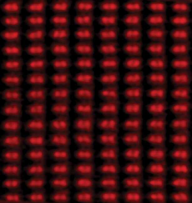
Electron microscopes
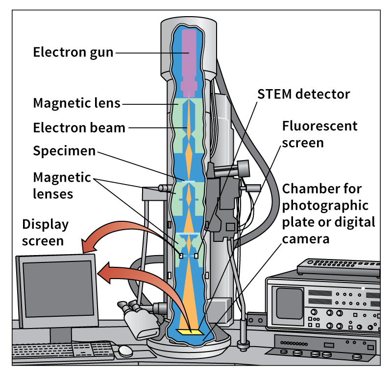
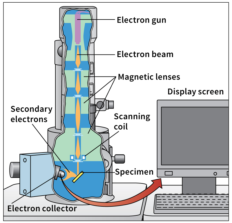
use a beam of electrons—rather than a beam of light—to produce magnified images. The wavelengths of electrons are much shorter than those of visible light. Some electron microscopes can resolve details that are smaller than 0.1 nanometer. This resolution is sufficient to reveal a sample’s fundamental atomic structure. However, electron microscopes cannot be used on living samples, in part because the electrons would damage cells.
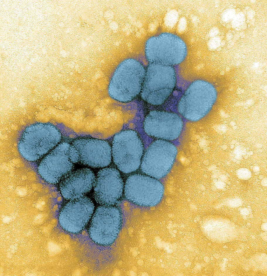
There are two basic types of electron microscope. A transmission electron microscope passes a broad beam of electrons through a sample slice a few dozen nanometers thick. A scanning electron microscope scans a focused beam across the specimen’s surface, building an image point by point.
Scanning probe microscopes
scan a specimen with a specialized tip called a probe. There are two main types of scanning probe microscopes: (1) the scanning tunneling microscope (STM) and (2) the atomic force microscope (AFM).
In the STM, the probe does not quite touch the specimen. But it comes close enough that an electric current “tunnels,” or transfers, between the probe and the specimen. A computer uses measurements of this current to create an image. An STM can resolve surface atoms that are less than 0.1 nanometer apart. The STM is widely used in materials science to examine the surfaces of materials, such as computer chips.
In the AFM, the probe usually gently touches the specimen’s surface. As the probe scans the specimen, it reacts to the roughness of the surface by moving up and down. Electronic devices measure this movement and send their results to a computer, which creates an image. The AFM can produce images of specimens, such as animal tissue, through which electric current does not flow readily. Biologists increasingly use the AFM to obtain high-resolution images of living samples.
Ion microscopes,
also known as field-ion microscopes, are used to examine metals. An ion microscope creates an image of the crystal structure of a metal that has been shaped into an extremely sharp needle. An electric field applied to the needle’s tip repels ions (charged atoms) of helium, neon, or argon. The ions spread out and strike a special screen. The screen glows where the ions strike it. The glows, in turn, form an image of the arrangement of atoms in the metal. Ion microscopes, like STM’s, are most commonly used in materials science.
History
Engravers probably used water-filled glass globes as magnifying glasses at least 2,000 years ago. The Romans may have made magnifying glasses from rock crystal. Modern glass lenses were introduced in the late 1200’s.
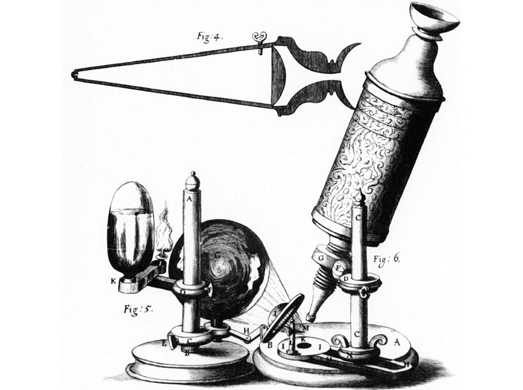
Historians generally credit a Dutch spectacle-maker, Zacharias Janssen, with discovering the principle of the compound microscope about 1590. In 1665, the English scientist Robert Hooke used an early microscope to discover plant cells in a slice of cork. He named the structures “cells” because they looked like little walled rooms.
In the 1670’s, Anton van Leeuwenhoek, a Dutch amateur scientist, made single-lens microscopes that could magnify up to 270X. He was the first person to observe microscopic life and record his observations. He is therefore considered the father of modern microscopy.
Few improvements in microscopes occurred until the early 1800’s. At that time, improved glassmaking methods produced lenses that provided undistorted images.
German scientists demonstrated the first electron microscope in 1931. The ion microscope was invented in 1951. In 1981, Swiss and West German scientists demonstrated the first scanning tunneling microscope.
Modern research in microscopy largely involves improving the resolution and sensitivity of optical microscopes. Advances in computers and engineering have enabled researchers to create a special type of optical microscope that can show details as small as about 20 nanometers. Such microscopes use specialized techniques to produce images with resolutions much finer than the wavelength of visible light. These microscopes can even detect individual molecules in living cells.

