Nervous system is an internal communications network that enables an animal to adjust to changes in its environment. Almost all animals, except the simplest kinds, have some type of nervous system.
Animals without a backbone have a nervous system that ranges from a simple net of nerves to a highly organized system of nerve cords and a primitive brain. In human beings and other animals with backbones, the nervous system consists of the brain, the spinal cord, and the nerves. This article deals mainly with the human nervous system.
The human nervous system—especially the highly developed brain—makes people different from all other animals. The human brain functions much like a complicated computer that enables people to speak, solve difficult problems, and produce creative ideas.
The nervous system provides pathways by which information travels from a person’s surroundings to the brain. The brain then sends instructions to various muscles via other pathways so that the body can respond to the information. The nervous system also regulates internal functions, such as breathing, digestion, and heartbeat. All of a person’s movements, sensations, thoughts, and emotions are products of his or her nervous system.
How the nervous system works
The nervous system is made up of billions of special cells called neurons or nerve cells. Cordlike bundles of neuron fibers are called nerves. The nerves form a network of pathways that conduct information rapidly throughout the body.
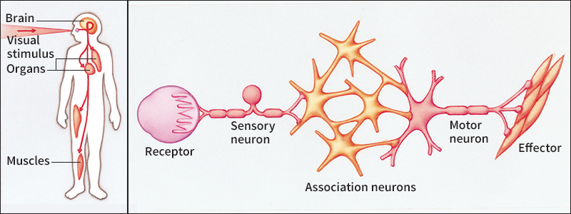
A person’s reaction to a situation may take only an instant, but it involves many complicated processes within the nervous system. For example, what happens in the nervous system of a person who sees a wild tiger and, an instant later, turns and runs away?
Specialized neurons called receptors are located in the ears and eyes and the other sense organs of the body. The receptors translate events in a person’s surroundings—such as the sight of a tiger—into nerve messages, which are known as impulses. Nerve impulses travel along nerve fibers at speeds of 3 to 300 feet (0.9 to 90 meters) per second.
The receptor cells in the eyes respond to light rays that reflect off the tiger and translate the rays into a pattern of nerve impulses. These impulses then travel through neurons called sensory neurons and association neurons. The sensory neurons carry information from receptors in the sense organs to the association neurons, which are located in the brain and the spinal cord.
The neurons in the brain receive the impulses, analyze and interpret the message, and decide what action should be taken. A message consisting of the sight of a wild tiger is, of course, interpreted as danger. The person’s brain immediately sends out a message—”Run!”—in the form of nerve impulses.
Next, the impulses travel through motor neurons. These nerve cells carry messages from the brain to the muscles and glands, which are called effectors. The effectors carry out the brain’s instructions. Thus, the leg muscles respond and the person runs away. At the same time, the brain sends messages to various other parts of the body. For example, it sends messages to the heart to beat faster and send more blood to the leg muscles.
Divisions of the nervous system
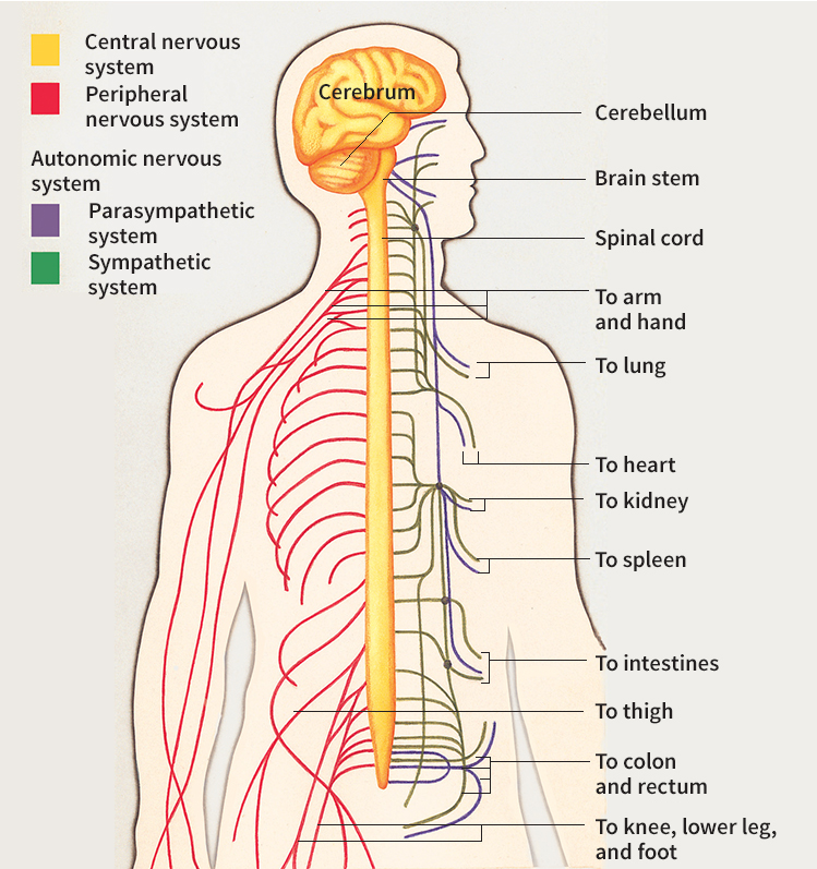
The nervous system has three main parts, the central nervous system, the peripheral nervous system, and the autonomic nervous system. Each of these parts has special functions.
The central nervous system
functions as a “main switchboard” that controls and coordinates the activities of the entire nervous system. The central nervous system consists of the brain and the spinal cord.
The brain is an extremely complicated organ. It consists of three principal parts, the cerebrum, the cerebellum, and the brain stem. This article provides basic information about the brain. For more detailed information on the brain and how it works, see Brain.
The cerebrum makes up about 85 per cent of the brain and is the most complex part. It is above the cerebellum and the brain stem and almost surrounds them. Human beings have a highly developed cerebrum that directs their hearing, sight, and touch and their ability to think, use language, and feel emotions. The cerebrum is also the center of learning.
The cerebellum, which is about the size of an orange, is slightly above the brain stem. It helps maintain the body’s sense of balance and coordinates muscular movements with sensory information.
The brain stem is a stalklike structure that is connected to the spinal cord at the base of the skull. The brain stem contains neurons that relay information from the sense organs. Many neurons that regulate automatic functions, such as balance, blood pressure, breathing, and heartbeat, are also in the brain stem.
The spinal cord is a cable of neurons that extends from the neck about two-thirds of the way down the backbone. The backbone surrounds and protects the spinal cord. The spinal cord contains pathways that carry sensory information to the brain. It also has pathways that relay commands from the brain to the motor neurons.
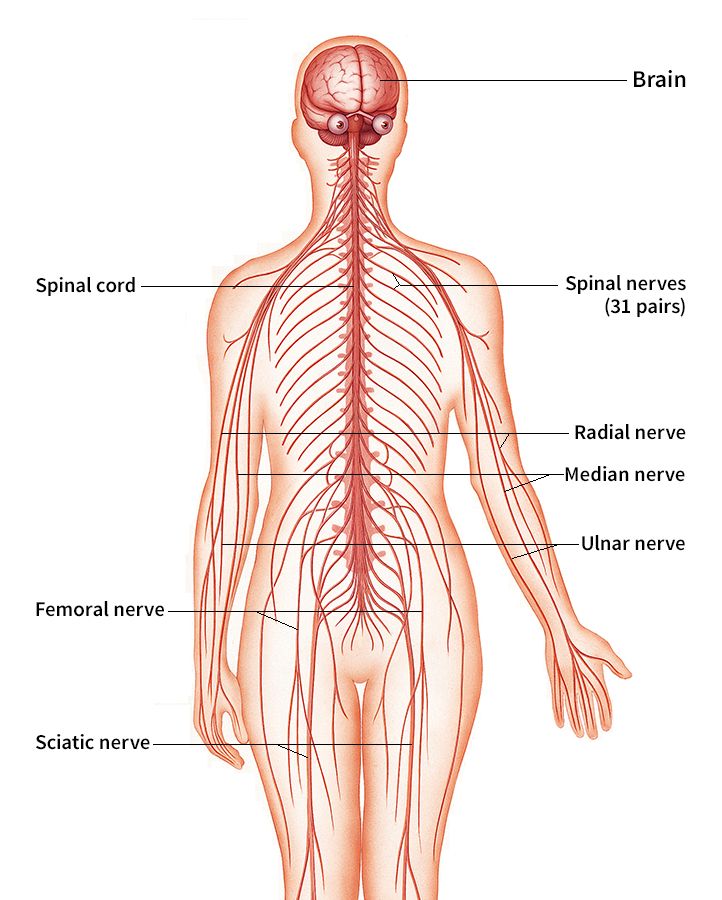
The peripheral nervous system
carries all the messages sent between the central nervous system and the rest of the body. The peripheral nervous system consists of 12 pairs of nerves that originate in the brain, plus 31 pairs of nerves of the spinal cord. These cranial and spinal nerves serve as “telephone wires” that carry messages to and from every receptor and effector in the body.
The autonomic nervous system
is a special part of the peripheral nervous system. The autonomic nervous system regulates such automatic bodily processes as breathing and digestion without conscious control by the brain. This constant regulation enables the body to maintain a stable internal environment.
The autonomic nervous system has two parts, the sympathetic system and the parasympathetic system. The sympathetic system responds to the body’s needs during increased activity and in emergencies. The actions of the sympathetic system include speeding up the heartbeat, sending additional blood to the muscles, and enlarging the pupils of the eyes to use all available light.
The parasympathetic system, in general, opposes the actions of the sympathetic system. The parasympathetic system’s functions include slowing down the heartbeat, diverting blood from the muscles to the stomach and intestines, and contracting the pupils of the eyes. The balance of activity between the two systems is controlled by the central nervous system.
Parts of a neuron
A neuron has three basic parts, the cell body, the axon, and the dendrites. A thin nerve membrane surrounds the entire cell.
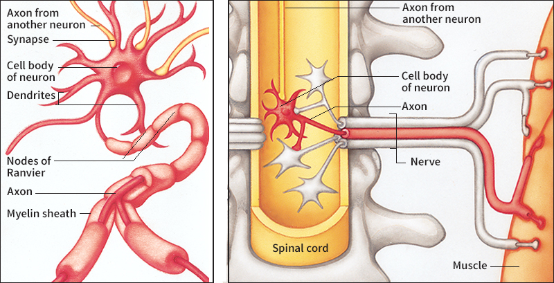
The cell body
of a neuron is a ball-shaped structure about 1/1000 of an inch (. 025 millimeter) wide. Each neuron cell body is a center for receiving and sending nerve impulses. The cell body is also responsible for making proteins and using energy for the maintenance and growth of the nerve cell.
The vast majority of neuron cell bodies are within the central nervous system, where incoming messages are combined and outgoing messages are produced. The few neuron cell bodies outside the central nervous system are grouped into clusters called ganglia. The best-known ganglia are in the autonomic nervous system.
The axon,
also called the nerve fiber, is a tubelike extension of a neuron cell body. The axon is specialized to carry messages. An axon of one neuron may have enough branches to make contact with as many as 1,000 other neurons.
Most axons in the central nervous system are less than 1/25 of an inch (1 millimeter) long. However, many axons in the peripheral nervous system are longer, and some are much longer. For example, the axons that extend from the spinal cord to the muscles in the feet may be 30 to 40 inches (76 to 100 centimeters) long.
The structures commonly called nerves are actually bundles of axons lying next to one another in a cordlike formation. Nerves can be made up of the axons of motor neurons or sensory neurons, or of both.
Some axons are covered by a sheath of a white, fatty substance called myelin. The myelin increases the speed of impulses along the axons. Myelin also causes the distinction between the gray matter and white matter in the nervous system. Gray matter consists largely of unmyelinated axons (axons without myelin sheaths) and neuron cell bodies. White matter is made up mostly of axons that have white sheaths of myelin. Myelin is formed by special supporting cells called glia that surround the neuron cell bodies and axons. In the central nervous system, glial cells called oligodendrocytes produce myelin. In the peripheral nervous system, the myelin that surrounds the axons is produced by glia called Schwann cells.
The dendrites
of a neuron are branching, tubelike extensions of the cell body that form a pattern resembling the limbs of a tree. Most neuron cell bodies have about six main dendrites, each of which is two or three times as thick as the axon of the cell. The distance between the cell body and the tips of the dendrites is about 1/50 of an inch (0.5 millimeter).
Dendrites are specialized structures for receiving impulses, mostly from the axon of another neuron. Dendrites and axons do not quite touch each other. In almost all cases, they are separated by an extremely narrow space called the synaptic cleft, over which nerve impulses are transmitted. These places where one neuron communicates with another are called synapses.
How messages are routed
Simple pathways.
Much of the work of the nervous system depends upon established pathways between neurons. These pathways are called neural circuits. The simplest kind of neural circuit is a reflex. A reflex is an automatic and involuntary response to a stimulus. Simple reflexes do not involve the brain. Impulses in a reflex action follow a simple pathway that goes through the spinal cord and links a receptor and an effector.
One of the simplest reflexes is the knee jerk reflex. A tap on the tendon below the kneecap stretches the muscle there, and this action stimulates special receptors to produce an impulse. The impulse travels through a sensory neuron, over an axon to the spinal cord, and through a synapse to a motor neuron. There, a second impulse is generated. The second impulse travels over the axon of a motor neuron back to the muscle that was stretched. The impulse causes the muscle cells to contract and the leg to jerk.
Complex pathways.
Many reflexes are more complicated. They involve at least one association neuron between the sensory and motor neurons. The association neuron, in turn, may be connected with many complex neural pathways, some of which may lead to the brain. A common but complex reflex is involved in withdrawing from a painful stimulus. For example, if a person steps on a sharp object, the immediate response is to lift the foot. In addition, association neurons stimulate the muscles of the other leg to adjust and maintain balance. Interconnecting neural pathways to the brain also may be stimulated so that the person becomes aware of what has happened.
Reflexes alone cannot account for the vast number and variety of actions performed by human beings. People, as well as some other kinds of animals, can learn new behavior patterns. Voluntary muscular movements necessary for learning new skills travel along complex nerve pathways that extend from the brain throughout the body. Complicated actions, such as riding a bicycle or walking, eventually can be performed without constant, conscious control after they have been learned.
How neurons carry impulses
During the 1800’s, scientists discovered that nerve impulses involve electrical charges. They assumed a nerve impulse was simply an electric current flowing through nerves. By the 1900’s, researchers had learned that different concentrations of certain ions in neurons and in their surrounding fluids create a potential electrical charge. It was also learned that nerve cell membranes have pores that allow only certain substances to pass through. Scientists then theorized that a nerve impulse was an electrochemical process controlled by the nerve cell membrane.
In the 1930’s, researchers developed techniques to test the membrane theory of nerve conduction, which is the theory discussed in this section. This theory is the accepted explanation of how neurons carry impulses.
The beginning of an impulse.
A nerve cell membrane has special protein molecules that control the opening and closing of its pores. When at rest, the membrane keeps the concentration of sodium ions in the neuron very low. The membrane also keeps the concentration of potassium ions and negative organic ions much higher in the cell than in the surrounding fluids. These differences in ion concentration make the inside of the neuron more negative than the outside, and so the membrane is said to be polarized. The resulting voltage difference across the membrane is called the resting potential.
A chemical, electrical, or mechanical stimulus applied to a neuron can affect the membrane’s porosity and change the resting potential. The stimulus can cause the membrane’s pores to open and allow more sodium ions into the cell. The increase in sodium ions makes the inside of the cell positively charged, and this voltage change is called a depolarization.
When a stimulus causes a neuron to depolarize, the neuron is said to fire. The firing is the start of a nerve impulse. A stimulus must be of a certain intensity, called the threshold voltage, for a neuron to fire.
All impulses from a particular neuron have the same size and duration, no matter how large the stimulus that caused the neuron to fire. The fact that neurons fire at maximum strength or not at all is called the all-or-nothing phenomenon. The brain probably detects the intensity of a stimulus by the frequency of impulses generated and the number of nerve fibers stimulated.
Conduction along the axon.
The inside of an axon is filled with a solution that can conduct an electric charge. Depolarizations in one area of an axon spread through the solution to neighboring areas along the axon. This wave of depolarizations is called an action potential.
If the axon of a neuron has no myelin sheath, the nerve impulse sweeps continuously along the axon, like fire along a firecracker fuse. But in a myelinated axon, nerve impulses can occur only at the nodes of Ranvier. The nodes of Ranvier are areas along an axon where the myelin sheath is interrupted at regular intervals. The impulse hops from node to node.
Transmission across synapses.
Certain chemicals, called neurotransmitters, transmit nerve impulses across synapses. When an impulse reaches the end of an axon, a neurotransmitter is released into the synaptic cleft. The neurotransmitter moves to the dendrites of the next nerve cell and causes certain pores of the nerve membrane to open. Ions move through these pores, and a voltage change, called a postsynaptic potential, results.
The postsynaptic potential is either excitatory or inhibitory. An excitatory postsynaptic potential spreads to the axon of a nerve cell and tends to produce another action potential. An inhibitory postsynaptic potential tends to prevent the axon from producing another action potential. Not every impulse that reaches a synapse is transmitted to the next neuron. The synapses thus help regulate and route the constant flow of nerve impulses throughout the nervous system.
Disorders of the nervous system
The nervous system can be damaged by injury and disease. Axons in the central nervous system cannot regrow after being damaged, but nerves in the peripheral nervous system may recover. Severely damaged nerve cells that die cannot be replaced.
Most neurons that perform a specific job are grouped together in the brain. Because of this grouping arrangement, called localization of function, damage to one area of the brain may affect only certain abilities and leave others intact. In some cases, undamaged areas of the brain gradually assume control of functions lost when another area of the brain was damaged. This action is called recovery of function.
The most common serious disorder of the nervous system is stroke. A stroke occurs if the blood supply to a certain area of the brain is cut off, resulting in the death of nerve cells. Stroke victims may lose the ability to perform functions controlled by the damaged area of the brain, such as speaking or moving a limb. Stroke victims may eventually recover some lost functions. But if respiration or some other vital function is affected, a stroke can be fatal.
The most common infectious diseases that affect the nervous system are mild virus infections that last only a few days and may produce headaches. More serious infectious diseases, such as encephalitis and meningitis, are caused by certain bacteria, viruses, or other microbes. Encephalitis is an inflammation of the brain, and meningitis is an inflammation of the membranes covering the brain and the spinal cord.
The cause of multiple sclerosis, a disease of the nervous system, is not known. Multiple sclerosis causes axons in various areas of the central nervous system to lose their myelin sheaths. As a result, these axons cannot conduct nerve impulses properly.
Another disorder of the nervous system is epilepsy. Victims of epilepsy suffer seizures that can cause muscle convulsions and a change in, or loss of, consciousness. An epileptic seizure occurs if most of the neurons in one area of the brain produce bursts of impulses at the same time. Physicians prescribe drugs to reduce the number of seizures or to prevent seizures from occurring at all.
Before the development of vaccines to prevent poliomyelitis, this virus disease of the nervous system was widespread. The polio virus can destroy motor neurons in the spinal cord and brain stem, leading to paralysis in some cases.
Some disorders of the nervous system can lead to mental illness or intellectual disability. For information about these conditions, see the articles on Mental illness and Intellectual disability.
The nervous system in other animals
In vertebrates.
All vertebrates, including other mammals, as well as amphibians, birds, fish, and reptiles, have nervous systems much like the human nervous system. The bodies of the nerve cells that make up the nervous systems of these animals are about the same size and shape as those of human nerve cells.
The size of a specific area of the brain may indicate the importance of the function of that area for the animal. For example, dogs have a larger and better developed area for smell than do human beings. In contrast, human beings have a larger and more highly developed cerebral cortex than other animals. The cerebral cortex is the outer surface of the cerebrum, where such complicated skills as delicate motor control and the use of language are coordinated.
In invertebrates.
Most species of invertebrates that consist of more than one cell have some sort of nervous system. Many of the neurons of these animals are larger than human neurons. In hydras and some other simple invertebrates, the nervous system may be a nerve net, in which nerve cells are spread throughout the organism. There is no distinction between axons and dendrites in nerve net systems, and impulses travel in all directions from the point of stimulation.
Other invertebrates, including worms and insects, have more complicated, centralized nervous systems. These systems consist largely of concentrations of neurons that form a nerve cord. Ganglia along the cord serve as centers for organizing and integrating various activities of the animals. Clusters of ganglia in the front end of the body act as a primitive “brain.” Many insects also have such ganglia in the thorax region. These ganglia coordinate motor activities. The mechanisms of nerve impulses and synaptic potentials in higher invertebrates are the same as those in the human nervous system.