Blood is the river of life that flows through the human body. We cannot live without it. The heart pumps blood to all our body cells, supplying them with oxygen and food. At the same time, blood carries carbon dioxide and other waste products from the cells. Blood also fights infection, keeps our temperature steady, and carries chemicals that regulate many body functions. Finally, blood even has substances that plug broken blood vessels and so prevent us from bleeding to death.

When oxygen combines with certain cells—the red blood cells—the blood takes on its characteristic red color. Thus, blood that escapes from the body through a broken vessel appears bright red because of the oxygen in the air. Blood carrying oxygen to body cells has that same brilliant red color. But it turns a dark brownish-red after delivering oxygen.
The amount of blood in your body depends on your size and the altitude at which you live. An adult who weighs 160 pounds (73 kilograms) has about 5 quarts (4.7 liters) of blood. An 80-pound (36-kilogram) child has about half that amount, and an 8-pound (3.6-kilogram) infant has about 81/2 ounces (250 milliliters). People who live at high altitudes, where less oxygen is available, may have up to 2 quarts (1.9 liters) more blood than people who live in low regions. The extra blood delivers additional oxygen to body cells.
Blood also circulates through the bodies of dogs, cats, birds, insects, and most other kinds of animals. Only such simple animals as jellyfish and sponges do not need blood to live. For information about blood in some types of animals, see Circulatory system (The circulatory system in other animals). See also Mammal (Internal organ systems); Insect (Circulatory system).
The composition of blood
Blood consists of cells that move about in a watery liquid called plasma. The cells are known as formed elements because they have definite shapes. Three types of cells make up the formed elements: (1) red blood cells, (2) white blood cells, and (3) platelets. A microliter (1/30,000 of an ounce) of blood normally contains about 4 million to 6 million red blood cells, 5,000 to 10,000 white blood cells, and 150,000 to 500,000 platelets. The red and white blood cells are also called corpuscles.
Plasma
is the liquid, straw-colored part of blood. It makes up about 50 to 60 percent of the total volume of blood. The formed elements account for the rest.
Plasma consists of about 90 percent water. Hundreds of other substances make up the balance. They include proteins that enable blood to clot and to fight infection; dissolved nutrients (foods); and waste products. Plasma also carries chemicals called hormones, which control growth and certain other body functions.
Red blood cells,
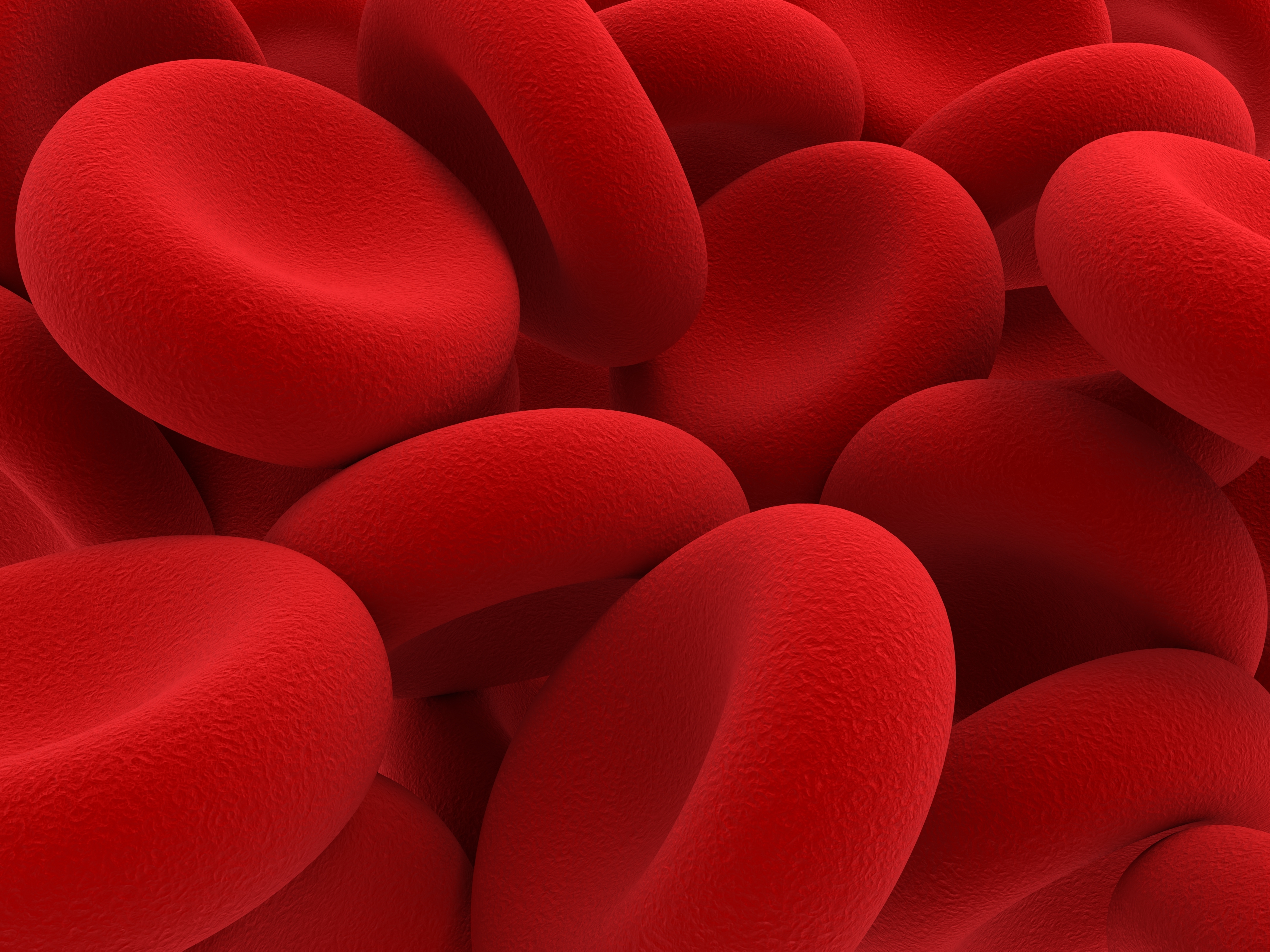
also called erythrocytes << ih RIHTH roh sytz >>, carry oxygen to body tissues and remove carbon dioxide. A red blood cell has a flat, disklike shape. It is thinner in the middle than at the edges—somewhat like a doughnut without the hole.
Red blood cells consist mainly of hemoglobin << HEE muh `gloh` buhn >>, an oxygen-carrying protein that gives them their red color. The cells also contain chemicals, particularly enzymes. Enzymes enable the cells to carry out necessary chemical processes more effectively. A flexible membrane surrounds each red blood cell. The membrane is so flexible the cells can squeeze through the tiniest blood vessels. Most kinds of cells have a nucleus, a central structure that controls many cell activities. But mature red blood cells have no nuclei.
White blood cells,
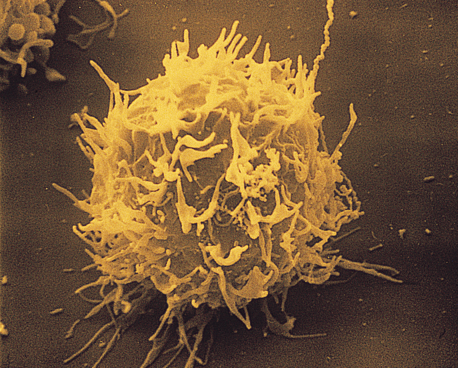
also called leukocytes << LOO kuh sytz >>, fight infections and harmful substances that invade the body. Most of the cells are round and colorless. They have several sizes, and their nuclei vary in shape. Some kinds of white blood cells kill bacteria by surrounding and digesting them. Other kinds produce antibodies, proteins that destroy bacteria, viruses, and other invaders or make them harmless.

Platelets,
also known as thrombocytes << THROM buh sytz >>, are disklike structures that help stop bleeding. They are the smallest formed elements. If a blood vessel is cut, platelets << PLAYT lihtz >> stick to the edges of the cut and to one another, forming a plug. They then release chemicals that react with fibrinogen << fy BRIHN uh juhn >> and certain other plasma proteins, leading to the formation of a blood clot.
What blood does in the body
The major jobs of blood are to transport oxygen and nutrients to body tissues and to remove wastes. To accomplish those tasks, blood must flow to all parts of the body. It does so by means of our circulatory system, which consists of the heart, a vast network of blood vessels, and the blood itself.
The heart pumps blood to all the body tissues. Blood leaves the heart through arteries and returns through veins. Within the tissues, the arteries become smaller and smaller. The smallest blood vessels are the capillaries. They connect the tiniest arteries and the tiniest veins. Oxygen, food, and other substances pass from the blood through the thin capillary walls into the tissues. Carbon dioxide and other wastes from the tissues also pass through the capillary walls and enter the bloodstream. Blood returns to the heart through ever-larger veins. For more information about how blood moves through the body, see Circulatory system; Heart.
Carrying oxygen and carbon dioxide.
All living cells in your body continuously absorb oxygen and give off carbon dioxide. Oxygen is carried to your body tissues mainly by hemoglobin in the red blood cells. Each molecule of hemoglobin binds easily with four molecules of oxygen. When you inhale, air enters the alveoli (air sacs) of your lungs. Oxygen from the air passes through the walls of the capillaries that surround each alveolus and binds with hemoglobin. Some oxygen also dissolves in the plasma. The bonds that hold hemoglobin and oxygen molecules together react to oxygen levels in the cells. If the oxygen levels are low, the bonds break easily, releasing oxygen.
Your cells use oxygen to produce energy. The process creates carbon dioxide, which passes from the cells through the capillary walls. Most carbon dioxide enters the plasma, but some attaches to hemoglobin. When the blood reaches the capillaries in your lungs, the carbon dioxide enters the alveoli and is exhaled. See Hemoglobin; Lung; Respiration (Gas transport between the lungs and tissues).
Transporting nutrients and wastes.
Food reaches your body tissues by means of the blood. After food passes through your stomach, it enters the small intestine, where digestion is completed. The wall of the small intestine has millions of tiny, fingerlike projections called villi. The villi absorb digested food molecules, which enter the capillary network of each villus and pass into the blood. Many nutrients bind with the plasma protein albumin, which carries them to body tissues.
Your cells use nutrients to produce the energy needed for cell growth, reproduction, and other functions. In producing energy, the cells create waste products. Like nutrients, wastes enter the bloodstream through the capillary walls. Many wastes bind with albumin or dissolve in the plasma, which transports them to the liver. The liver filters wastes and other harmful substances from the blood. It converts some wastes into a compound called urea. The blood carries urea to the kidneys, which remove it in urine. See Digestive system; Intestine; Liver; Kidney.
Protecting against disease.
White blood cells play an important role in your immune system, which helps your body resist disease-causing substances. The invasion of a harmful substance activates the white blood cells. They then work to destroy it. Some proteins in the plasma also help fight disease. There are five main groups of white blood cells.
Three kinds of white blood cells attack and destroy germs, especially bacteria, in a process called phagocytosis << `fag` uh sy TOH sihs >> . In phagocytosis, a white blood cell surrounds a germ and then kills it with enzymes. Such white blood cells are called phagocytes.
Neutrophils << NOO truh fihlz >>are the most numerous phagocytes. They fight mainly bacterial infections. When bacteria invade the body, neutrophils leave the bloodstream and travel to the infected area. Monocytes << MON uh sytz >>, like neutrophils, leave the bloodstream and migrate to infected tissues, where they mature and become macrophages << MAK ruh fayj uhz >>. Macrophages not only kill germs but also destroy cancer-causing cells. In addition, they help begin antibody production. Eosinophils << `ee` uh SIHN uh fihlz >>, a rare third kind of phagocyte, defend the body against parasites.
Members of a fourth group of white blood cells, lymphocytes << LIHM fuh sytz >>, do not perform phagocytosis. Instead, they have a key part in the body’s immune system by recognizing and responding to specific viruses, bacteria, and other invaders. There are two major kinds of lymphocytes—B cells and T cells. B cells produce antibodies and release them into the plasma, where they circulate in the form of globulin proteins. Such proteins, especially gamma globulin, fight infection (see Globulin; Gamma globulin). T cells release substances that control B-cell activity. They also produce substances that activate monocytes and so help destroy harmful organisms.
The chief function of a fifth group of white blood cells, basophils << BAY suh fihlz >>, is uncertain. Like eosinophils, they are rare blood cells.
To learn more about how white blood cells help us fight disease, see Immune system.
Carrying hormones.
Organs called endocrine glands produce hormones and release them directly into the blood. The hormones enter the plasma and act as “chemical messengers.” When a hormone reaches a part of the body it regulates, it may affect growth, reproductive processes, how the body uses food, or some other function. See Hormone; Gland.
Distributing body heat.
All cell activities produce heat. But some cells, particularly those in muscles and glands, create more heat than others. The heat enters your bloodstream and travels throughout your body. Excess heat escapes through your skin. If blood did not distribute heat, some body areas would become extremely hot while others would remain extremely cold. Thus, blood circulation helps keep your body temperature steady and safe.
How the body maintains its blood supply
You cannot live without a proper supply of healthy blood. In addition, the amounts of the various blood components (parts) must change constantly as the needs of your body change. Substances called hematopoietic growth factors govern the production of the red cells, white cells, and platelets. Your body maintains its blood supply by (1) regulating the volume of blood components, (2) controlling bleeding, and (3) replacing worn-out blood components.
Regulating the volume of blood components.
The volume of each blood component continuously adjusts to meet the body’s needs. The plasma proteins, especially albumin, control the movement of plasma between the capillaries and the cells. Normally, only dissolved substances, such as nutrients, pass from the plasma through the capillary walls. But if the amount of albumin falls below normal, plasma may escape into tissues. In contrast, if the concentration of albumin is high, water from the tissues enters the plasma.
The volume of red blood cells depends on how much oxygen body tissues require. The kidneys produce a hormone called erythropoietin that stimulates output of the cells. When the tissues need oxygen, the kidneys produce increased amounts of erythropoietin, causing red-cell production to rise. When oxygen need falls, erythropoietin output drops. Certain diseases also may reduce the production of red blood cells.
Other hematopoietic growth factors control the number of white blood cells and platelets, which also increase and decrease according to the condition of the body. For example, an infection leads to a rise in the number of germ-fighting white blood cells. Similarly, severe bleeding can cause an increase in the number of platelets, thus improving the blood’s ability to clot.
Controlling bleeding.
You would bleed to death from a small cut if your blood did not coagulate (clot). An injured blood vessel causes platelets to stick to the damaged surface and to one another, forming a plug.
The plasma contains proteins called clotting factors. They normally circulate in an inactive form in the blood. But if a blood vessel suffers damage, the platelet plug and the injured vessel give off chemicals that react with the clotting factors. Eventually, the plasma protein fibrinogen changes into sticky strands of fibrin. The strands crisscross one another, creating a mesh that holds red blood cells and the platelet plug tightly to the site of bleeding. The fluid is squeezed out, and a solid plug—the clot—forms. A clot on the skin surface is a scab.
Occasionally, a clot may occur in an undamaged vessel that has no bleeding. Such a clot, called a thrombus, may block the flow of blood to tissues beyond the clot and cut off food and oxygen to those tissues. If a clot blocks an artery that nourishes the heart, a coronary thrombosis results, which may cause a heart attack (see Coronary thrombosis). If a clot blocks an artery to the brain, a stroke may occur (see Stroke).
Blood contains substances that dissolve clots as well as produce them. These substances circulate in an inactive form until clotting occurs. They are then activated to control the extent and duration of the clotting.
Replacing worn-out blood components.
Each formed element can live only a particular length of time, and so your body must continuously replace worn-out cells. Red blood cells live about 120 days, and platelets about 10 days. The life span of white blood cells varies greatly. For example, neutrophils live only a few hours, dying soon after they perform phagocytosis. But some lymphocytes live many years, thus providing long-term immunity against certain diseases.
Destruction of worn-out blood components.
Two body organs—the liver and the spleen—remove worn-out red blood cells from the bloodstream and break them down. The liver uses coloring matter from the old cells in producing a digestive liquid called bile. The iron from hemoglobin is reused by the body to make new red blood cells. Worn-out white blood cells migrate to body tissues, where they die. Platelets probably wear out plugging tiny leaks in blood vessels.
Formation of new blood components.
The core of human bones is filled with a soft red or yellow substance called marrow. In adults, the red bone marrow produces millions of blood cells per second. Red marrow occurs mostly in flat bones, such as the vertebrae, ribs, and skull. All blood cells begin in the marrow as stem cells. They develop into more mature precursor cells for each type of blood cell, each of which forms either many red blood cells, white blood cells, or platelets.
As red blood cells develop in the marrow, they make hemoglobin. They also shrink and lose their nuclei. At maturity, they enter the bloodstream through tiny blood-filled cavities, called sinuses, in the marrow.
Although all white blood cells originate in the red bone marrow, lymphocytes—the T cells and B cells—mature elsewhere in the body. T cells enter the bloodstream through the sinuses and move to the thymus, a gland near the base of the neck, where they complete their development. The mature T cells then travel to structures called lymph nodes, which occur in many areas of the body. B cells complete their maturation in the lymph nodes and spleen.
Platelets develop in the red marrow from large precursor cells called megakaryocytes << `mehg` uh KAR ee uh sytz >>. They eventually split into fragments, each of which becomes a platelet and enters the bloodstream.
Blood groups
The membranes of red blood cells contain proteins called antigens. More than 300 red-cell antigens have been identified. Based on the presence or absence of particular antigens, scientists have classified human blood into various groups.
The significance of blood groups.
Blood-group classifications have extreme importance in certain medical procedures. Information about blood groups has also been used in law and anthropology.
In medicine,
the chief use of blood groups is to determine whether the blood of one person, called a donor, can be transfused into the body of a patient without rejection or serious reaction. In almost every person, the plasma contains antibodies that react to certain antigens not present on the surface of that person’s own red blood cells. During a transfusion, dangerous clumping of the red blood cells may occur if antibodies in the patient’s plasma bind to antigens on the donor’s red blood cells. The clumping can block small blood vessels and result in severe illness or even death. No one’s plasma normally contains antibodies that bind with the person’s own red-cell antigens.
The most serious transfusion reaction is the rapid destruction of the transfused red blood cells. This may lead to shock, kidney failure, and sometimes death. Other reactions may include fever, shaking, and chills.
Before a patient has a blood transfusion, hospitals always perform a cross-match, a test in which a sample of the donor’s red blood cells is mixed with a sample of the patient’s plasma. If clumping occurs, the patient does not receive blood from that donor. Cross-matching thus reduces the possibility of dangerous transfusion reactions.
The membranes of white blood cells carry proteins called HLA antigens. Physicians use the presence of those antigens to help determine whether an organ or tissue from a certain donor can be safely transplanted into a patient (see Transplant).
In law.
In the past, law enforcement officials have used blood groups to help uncover the identity of criminals. For example, a blood specimen from the scene of a crime can be compared with that of a suspect. Today, DNA analysis is used to more accurately identify blood specimens.
The antigens on red blood cells are inherited, and so blood tests have been used in paternity cases, in which a man is accused of being a child’s father. The tests cannot prove that a certain man fathered a certain child, but they can sometimes prove that he did not. The use of blood groups in paternity and other parenthood cases has been largely replaced by studies of the DNA molecules in blood cells. DNA carries hereditary information in all the body cells, and such tests are almost 100 percent accurate in determining parenthood.
In anthropology.
In the past, anthropologists studied the relative frequencies of blood groups among human populations. They believed that differences in blood group frequencies may reflect differences among human races. But most anthropologists today think that racial classification based on such physical characteristics is not scientifically valid and serves no useful purpose.
The ABO blood groups
make up the leading system of blood classification. The system classifies human blood into four main types, or groups. The types are based on the presence or absence of two antigens, called A and B, on the surface of red blood cells. (1) If the cells have only antigen A, the blood is type A. The plasma contains anti-B antibodies, which clump cells having antigen B. (2) If the red cells have only antigen B, the blood is type B. The plasma contains anti-A antibodies, which clump cells having antigen A. (3) If the cells have both antigens A and B, the blood is type AB. The plasma contains neither anti-A nor anti-B antibodies. (4) If the red cells have neither antigen A nor antigen B, the blood is type O. The plasma contains both anti-A and anti-B antibodies. Worldwide, type O blood is the most common, followed by type A. Relatively few people have type B, and even fewer have type AB.
Doctors prefer to use donor blood of the same ABO type as that of the patient to avoid clumping during a transfusion. But in an emergency, type O blood may be transfused into patients of any blood type. Similarly, type AB patients may be able to receive any ABO blood in an emergency because they have no antibodies to A or B antigens. But even then, hospitals perform a cross-match to ensure that no clumping will occur. Type A patients should never receive type B blood, and type B patients should never receive type A blood.
In most cases, it does not matter if the donor’s plasma contains antibodies that clump the patient’s red blood cells. The plasma dilutes rapidly in the patient’s blood, making the risk of clumping slight.
Rh blood types
form the second major blood-group system. People who have Rh antigens on their red blood cells are Rh positive. The antigen itself is called the Rh factor. People who lack the factor are Rh negative. Most people are Rh positive.
Plasma has no natural antibody to the Rh antigen. But Rh-negative people may build up antibodies called anti-Rh if they receive a transfusion of Rh-positive blood. The donor blood usually dilutes quickly, and so the antibodies create no problems. But clumping will occur later if an Rh-negative patient receives another transfusion of Rh-positive blood, which causes the anti-Rh to attack the Rh-positive blood. A mixing of Rh-negative and Rh-positive blood can also happen if an Rh-negative woman becomes pregnant with an Rh-positive baby. If some of the baby’s red blood cells enter the woman’s blood, anti-Rh may build up in her plasma. The situation can cause serious problems if the mother later becomes pregnant with another Rh-positive baby. See Rh factor.
Other blood groups.
Many other systems for classifying blood have been developed. They include the Duffy, Kell, Kidd, Lewis, Lutheran, MNS, and P systems. But natural antibodies to the antigens in those systems occur rarely. Aside from the A and B antigens of the ABO system and the Rh factor, most red-cell antigens do not produce strong or dangerous reactions.
Medical uses of blood
Blood transfusions.
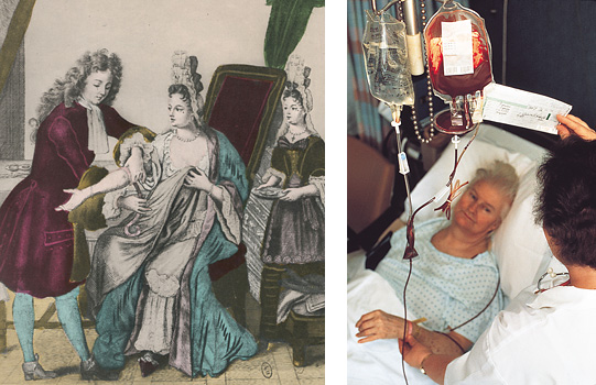
The ability to transfuse blood or blood components into sick or injured people has saved countless lives and revolutionized patient care. If an adult suddenly loses more than 1 quart (0.95 liter) of blood, death may occur unless the person receives a transfusion. Transfusions can also help patients whose bone marrow does not produce enough blood cells. In addition, transfusions replace blood lost during surgery.
Blood banks collect blood from donors and store it in sterile bags with a preservative and a chemical to help prevent clotting. Generally, patients need only one blood component, such as red blood cells. For that reason, blood banks separate most whole blood into components before storage. Whole blood can be refrigerated and stored for 21 to 49 days. Plasma, red blood cells, and certain other components can be frozen and stored up to several years.
Some diseases may be transmitted from a donor to a patient through a transfusion. Laboratory workers therefore screen all donated blood for the presence of hepatitis, AIDS, and certain other infectious diseases. In addition, a cross-match must ensure that no dangerous reactions will result. See Blood transfusion.
Blood tests.
Doctors use two main types of blood tests: (1) screening tests and (2) diagnostic tests.
Screening tests
help physicians detect unsuspected problems in patients. For example, a blood count calculates the number of red and white cells and the amount of hemoglobin in a sample of blood (see Blood count). A hematocrit measures the volume of red blood cells compared with other blood components. Abnormalities revealed by either test may indicate a disease or a defect in blood-cell production.
Doctors use various other blood tests to detect certain diseases. For instance, a test that shows a high level of glucose (sugar) in the blood may indicate diabetes, a disease in which the body does not use sugar normally (see Diabetes). A blood test that reveals a high level of the waste product urea may indicate a disorder of the kidneys, which filter urea from the blood. Physicians also screen patients’ blood for high levels of cholesterol, which has been associated with an increased risk of heart disease (see Cholesterol).
Diagnostic tests
help doctors discover the causes of some conditions. For example, anemia (an abnormally low number of red blood cells) may result if the diet does not include enough iron, vitamin B12, or folic acid (also called folate or folacin). The size of a patient’s red blood cells can reveal which nutrient the body needs. If the anemia results from too little iron, for example, the red cells are unusually small. But if it results from not enough vitamin B12, the cells are unusually large.
A differential white count test tells a doctor the percentage of each type of white blood cell in a patient’s blood. An extremely high number of white blood cells may mean leukemia, a form of cancer. On the other hand, a low neutrophil count may indicate an inability to fight infections effectively. Such diagnostic tests as a platelet count and a clotting test help physicians learn of certain bleeding disorders. The tests may also be performed before an operation to determine if the patient might bleed excessively during surgery.
Blood disorders
Disorders of the blood involve overproduction, underproduction, or excessive destruction of blood cells. Certain infections also can affect the blood.
Anemia
results from abnormally low levels of red blood cells or hemoglobin. A severely anemic person’s blood carries too little oxygen to meet the needs of body tissues.
Various conditions may cause anemia. One main cause is insufficient production of red blood cells by the bone marrow. The underproduction may stem from nutritional deficiency, disease, or infection. In addition, blood loss from an injury often results in anemia. Excessive hemolysis (destruction of red blood cells) may also cause anemia. Two hereditary diseases—sickle cell anemia and thalassemia—involve hemoglobin abnormalities. Physicians use diet therapy, drugs, transfusions, or a bone marrow transplant to treat anemia, depending on its cause.
White-cell abnormalities.
Acute leukemia results from uncontrolled and excessive white blood cell production. Physicians do not know exactly what causes the cancer. They use drugs, radiation, transfusions, or a bone marrow transplant to treat it.
The blood has an unusually low number of white blood cells in a disorder called leukopenia. It can result from exposure to certain drugs, diseases, or infections. In neutropenia, the most common type of leukopenia, the number of neutrophils is sharply reduced. People with neutropenia have an increased risk of infection because their blood lacks enough neutrophils to defend the body against harmful bacteria.
Bleeding disorders
come from a disruption of the blood’s ability to clot. Most such disorders result from abnormally low levels of clotting factors in the plasma or from an abnormality of the platelets.
A lack of some clotting factors causes hemophilia, a hereditary condition in which the blood coagulates extremely slowly. Hemophiliacs risk sudden, unexplained bleeding; severe bleeding from minor injuries; and bleeding of the joints and internal organs. Physicians treat the disorder by injecting the patient with the missing clotting factor.
Platelet abnormalities also affect the blood’s clotting ability. People with thrombocytopenia—that is, an unusually low number of platelets—risk dangerous episodes of bleeding. The low platelet count may be caused by certain drugs, infections, or increased platelet use by the body. People with thrombocythemia—that is, an excessive number of platelets—may also risk abnormal bleeding as well as abnormal clotting. A shortage of iron or the presence of cancer or certain other diseases may produce the high platelet count. Treating the causes of both conditions usually corrects them.
Infections.
Various infections can attack the blood. For example, infectious organisms can poison the blood and spread throughout the body. In malaria, a parasite destroys the red blood cells. In mononucleosis, a virus infects the B cells. In AIDS, a virus infects the T cells and damages the immune system.
History of blood research
Scientific interest in blood probably began with the Greek physician Hippocrates, who lived during the 400’s and 300’s B.C. He proposed that all diseases resulted from an imbalance of four humors (body fluids)—black bile, blood, phlegm, and yellow bile. The theory led to bloodletting—the drawing of blood from a vein of a sick person so the disease would flow out with the blood. For many centuries, bloodletting was standard medical treatment. Barbers performed the procedure during the Middle Ages. In the late 1700’s and early 1800’s, a number of doctors, especially the American physician Benjamin Rush, prescribed bloodletting to treat most illnesses. Some patients died of excessive blood loss.
In 1628, the English physician William Harvey described how blood circulates through the body. His work became the basis for later discoveries about the functions of blood. See Harvey, William.

In 1882, Elie Metchnikoff, a Russian biologist, discovered phagocytosis. His work helped explain how white blood cells kill germs. Also in 1882, an Italian biologist, Giulio Bizzozero, was the first to correctly describe the function of platelets and relate them to blood clotting.
As knowledge of blood components increased, interest in transfusions grew. Doctors first transfused blood directly from donors into patients. Most of the attempts failed. Then in the early 1900’s, Karl Landsteiner, an Austrian-born American physician, discovered the ABO blood types. Cross-matching blood types of donors and patients led to a dramatic increase in successful transfusions. In 1940, Landsteiner and Alexander S. Wiener, an American scientist, discovered the Rh factor.
The storage of blood became possible in 1914 with the addition of nutrients and of chemicals that checked clotting. In 1937, Bernard Fantus, an American physician, set up the first blood-bank program. Another American physician, Charles Drew, organized many such programs during World War II (1939-1945). Drew also urged the use of plasma, which at that time could be stored longer than whole blood, for battlefield and other emergency transfusions.
Scientists today are working to develop blood substitutes or artificial blood that could replace human blood in transfusions. Such research is very important because, even with strict precautions, transfusions involve risk of reactions and the transmission of viruses and other infections through transfused blood.
Other current research involves producing and testing the hematopoietic growth factors responsible for the formation of all blood cells. Many of the growth factors are available in large quantities for use in patients. They are being used in patients who lack enough red blood cells, white blood cells, or platelets.
