Ear is the sense organ that makes it possible for us to hear. Hearing is one of our most important senses. It enables us to communicate with one another through speech. The development of speech itself depends mostly on hearing. Children learn to talk by listening to and imitating the speech of other people. Hearing can also alert us to danger. We hear the warning honk of an automobile horn or the whistle of an approaching train. Even while asleep, we may hear a fire alarm or the barking of a watchdog. In addition, hearing provides pleasure. For example, it enables us to enjoy music, the singing of birds, and the sound of the surf.
Loading the player...Ear: Hearing
Hearing is a complicated process. Everything that moves makes a sound. Sound consists of vibrations that travel in waves. Sound waves enter the ear and are changed into nerve signals that are sent to the brain. The brain interprets the signals as sounds.
Besides enabling us to hear, our ears help us keep our balance. The ears have certain organs that respond to movements of the head. These organs inform the brain about any changes in the position of the head. The brain then sends messages to various muscles that keep our head and body steady as we stand, sit, walk, or move in any way.
Many kinds of animals have ears similar to those of human beings, and some have an extremely keen sense of hearing. Hearing is vital to the safety and survival of numerous animals. Sounds may warn them of approaching enemies or other dangers. In addition, many animals sing, growl, hiss, or make other sounds and depend partly on their sense of hearing to communicate with one another.
Parts of the ear
Human beings have an ear on each side of the head. The ears extend deep into the skull. Each ear has three main parts: (1) the outer ear, (2) the middle ear, and (3) the inner ear.
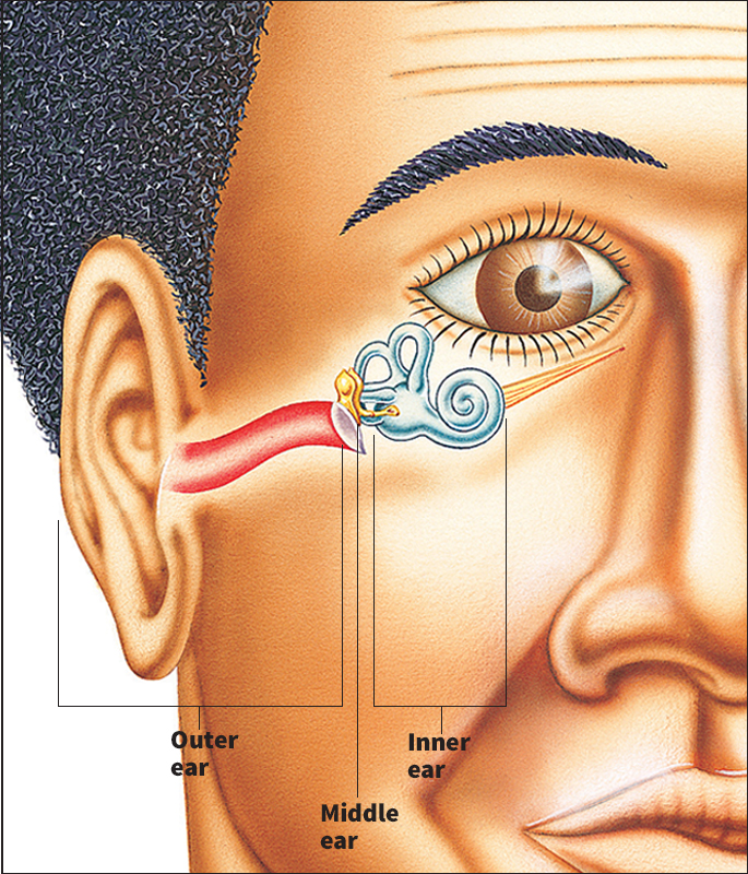
The outer ear
consists of three parts. They are: (1) the auricle, (2) the external auditory canal, and (3) the eardrum.
The auricle
is the fleshy, curved part of the ear on the outside of the head. The auricle has no bone. It consists mainly of tough, elastic tissue called cartilage, which is covered by a thin layer of skin. The loosely hanging lower part of the auricle is called the earlobe. It is made up of fat.
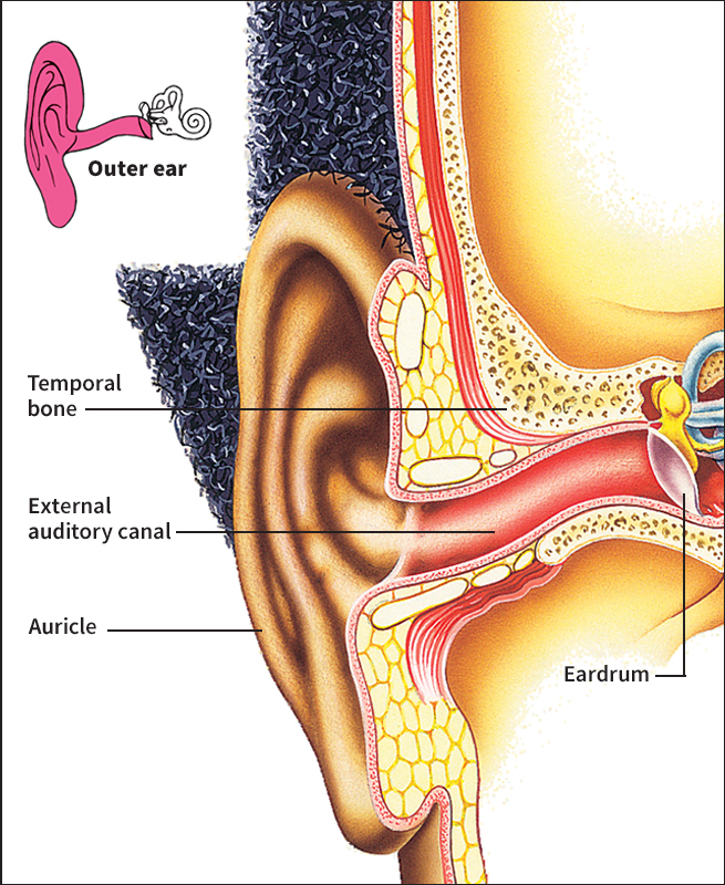
Three small muscles attach the auricle to the head. In human beings, these muscles have no practical use. However, some people can move them and so wiggle their ears. In many animals, these muscles are well developed and highly movable. Cats, dogs, foxes, horses, rabbits, and many other animals can turn their ears in the direction from which a sound is coming and so improve their hearing.
The external auditory canal
is the opening you see if you look directly into the ear. This passageway leads to the eardrum.
The external auditory canal is about 1 inch (2.5 centimeters) long. It curves somewhat in the shape of a C. The canal is lined with skin. The skin on the outer third of the canal has hairs, sweat glands, and glands that produce earwax. Earwax helps protect the eardrum by trapping dirt that would otherwise lodge against the membrane. Sometimes, earwax builds up in the canal and must be removed by a doctor. Never try to remove earwax yourself, especially by sticking small objects into the ear. You could easily puncture the eardrum.
The inner two-thirds of the auditory canal is surrounded by the temporal bone, which is the hardest bone in the body. The temporal bone also surrounds the middle ear and the inner ear. The bone protects the delicate structures of these parts of the ear.
The eardrum,
also called the tympanic membrane, separates the outer ear from the middle ear. It is a thin, round, tightly stretched membrane about 2/5 inch (10 millimeters) in diameter.
The middle ear
is a small chamber behind the eardrum. Three bones called the auditory ossicles extend across the chamber. The bones are linked together and connect the eardrum to the inner ear. The three bones have the Latin names malleus, which means hammer; incus, which means anvil; and stapes, which means stirrup. The bones look somewhat like the objects for which they are named. The malleus is the largest auditory ossicle. One end of the bone is attached to the eardrum, and the other end is joined to the incus. The incus, the second largest ossicle, connects the malleus to the stapes. The stapes is the smallest bone in the body. It is tinier than a grain of rice. The footplate of the stapes is attached to a membrane called the oval window, which leads to the inner ear.
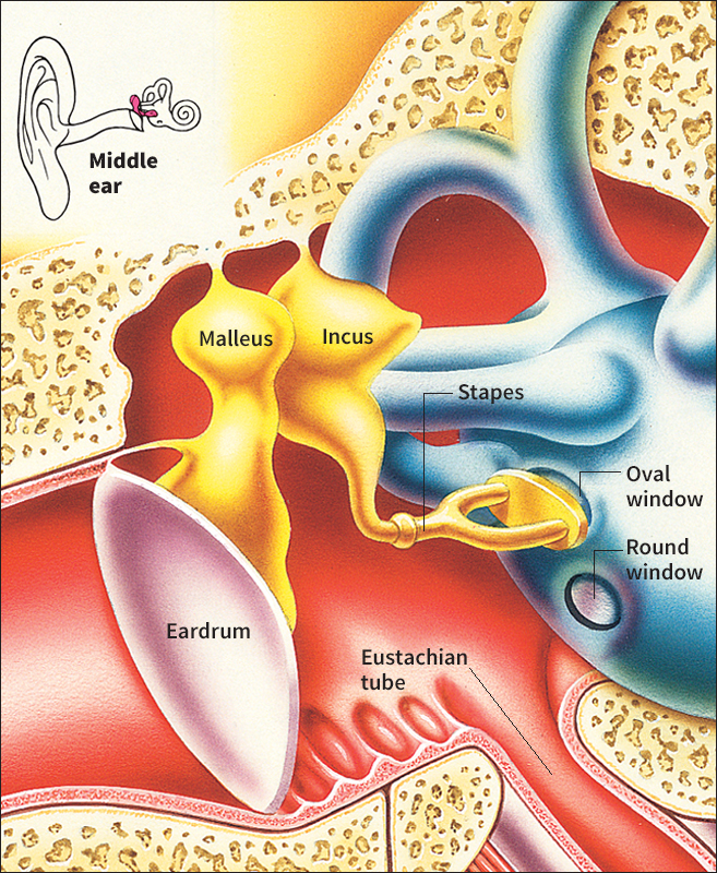
A narrow tube called the Eustachian tube connects the middle ear to the back of the throat. The tube is collapsed most of the time. It opens when you open your mouth, yawn, swallow, or blow your nose. When the Eustachian tube opens, air passes between the middle ear and the throat and so makes the air pressure on the inner side of the eardrum equal that on the outer side. If the Eustachian tube did not open, the eardrum would rupture when the air pressure, which varies with altitude, changes suddenly. The air pressure outside the eardrum changes rapidly, for example, when you go up or down quickly in an elevator, dive underwater, or land or take off in an airplane. In such cases, you have the sensation of “popping” of the ears as the Eustachian tube opens and allows air to escape from or to enter the middle ear.
The inner ear
has many delicate, interconnected structures and is sometimes called the labyrinth. A labyrinth is a group of passageways with a complicated arrangement. The inner ear consists of a bony labyrinth that encloses a narrower membranous labyrinth. A special kind of fluid is contained within the membranous labyrinth.
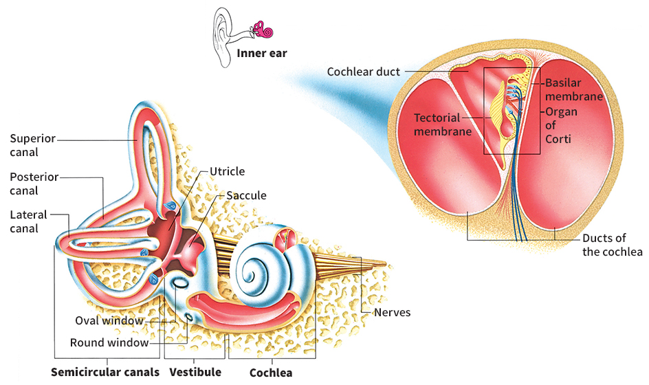
The inner ear has three basic parts, which are connected. They are (1) the vestibule, (2) the semicircular canals, and (3) the cochlea.
The vestibule
is a small, round chamber about 1/5 inch (5 millimeters) long. It forms the central part of the inner ear. Its bony walls connect the semicircular canals and the cochlea. Two sacs (baglike structures) lie within the vestibule. These sacs are called the utricle and the saccule. The inner wall of each sac has a swelling lined with hair cells. Hair cells are specialized sense cells with tiny, hairlike projections. The hair cells are attached to nerve fibers. A delicate membrane lies above the hair cells. Small mineral grains called otoliths are embedded in the membrane.
The vestibule has two small membranes that face the middle ear. One is the oval window, which is attached to the footplate of the stapes. The other is the round window, which lies just below the oval window.
The semicircular canals
are behind the vestibule. They consist of three canals set at right angles to one another. They are called the lateral, superior, and posterior canals. The lateral canal is horizontal. The two other canals are vertical. The superior canal lies in front of the posterior canal. Each canal forms two-thirds of a circle.
Each semicircular canal contains a fluid-filled duct (tube). One end of each duct widens and forms a pouch. This pouch, called the ampulla, has hair cells that are attached to nerve fibers.
The ducts of the semicircular canals are joined to the utricle. The utricle, in turn, is connected by a duct to the saccule. The semicircular canals and the utricle and saccule make up the inner ear’s organs of balance. They are sometimes called the vestibular organs or the labyrinthine organs.
The cochlea
is in front of the vestibule. It resembles a snail shell and forms a spiral that coils 21/2 times around its tip. Three fluid-filled ducts wind through the cochlea. One begins at the oval window, and another at the round window. These two ducts join at the tip of the spiral. The third duct, called the cochlear duct, lies between the first two. One wall of the cochlear duct consists of the basilar membrane. This membrane supports the organ of Corti, which has over 15,000 hair cells and is the actual organ of hearing. A membrane called the tectorial membrane lies above the hair cells.
The nerve of the inner ear is known as the auditory nerve or the vestibulocochlear nerve. It has two branches—the cochlear nerve and the vestibular nerve. Fibers of the cochlear nerve extend to each hair cell of the organ of Corti. Some fibers of the vestibular nerve lead to the hair cells of the utricle and the saccule, and others extend to the hair cells of the ampulla of each semicircular canal.
The sense of hearing
Sound consists of vibrations that travel in waves through the air, the ground, or some other substance or surface. Sounds vary in frequency and intensity. Frequency is the number of vibrations produced per second and is measured in hertz. One vibration per second equals 1 hertz. A high-frequency sound has a high pitch, and a low-frequency sound has a low pitch. The full range of normal human hearing extends from 20 to 20,000 hertz. As a person grows older, however, the ability to hear high-frequency sounds decreases. Intensity is the amount of energy in a sound wave. It is measured in decibels. A person can barely hear a sound of zero decibels. Sounds above 140 decibels can be painful to the ears. In some cases, they may seriously damage the ears.
This section describes (1) how sounds travel to the inner ear and (2) how sounds reach the brain.
How sounds travel to the inner ear.
Sound waves enter the external auditory canal of the outer ear and strike the eardrum, causing it to vibrate. The vibrations from the eardrum then travel across the three auditory ossicles of the middle ear—from the malleus, which is attached to the eardrum, to the incus and then to the stapes. The footplate of the stapes vibrates within the oval window, which creates waves in the fluid that fills the ducts of the cochlea of the inner ear.
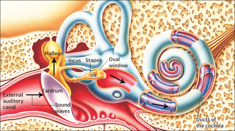
Besides transmitting sound waves to the oval window, the auditory ossicles of the middle ear amplify (strengthen) the waves. Sound waves do not travel as easily through the cochlear fluid of the inner ear as they do through the air. They diminish by about 30 decibels as they pass through the cochlear fluid. But amplification of the sound waves by the auditory ossicles makes up for the loss in intensity.
Sound waves can also be conducted to the inner ear through the bones of the skull. This process is called bone conduction. Some of the sound produced by your voice travels to your inner ears in this way. This fact explains why your own voice sounds different to you in a recording.
How sounds reach the brain.
The movements of the footplate of the stapes in the oval window produce waves in the cochlear fluid. The cochlear fluid pushes against the basilar membrane, causing it to move. The hair cells of the organ of Corti on the basilar membrane slide against the overhanging tectorial membrane. The hairs bend and so create impulses in the cochlear nerve fibers attached to the hair cells. The cochlear nerve transmits the impulses to the temporal lobe, the hearing center of the brain. The brain interprets the impulses as sounds. See Brain (Sensing the environment) .
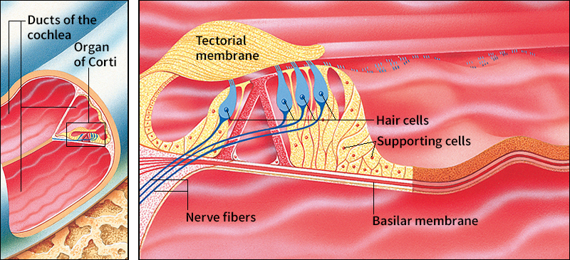
Sounds of high, middle, and low frequency affect hair cells at different locations along the basilar membrane. High-frequency sounds move hair cells near the base of the spiraling cochlea. Middle-frequency sounds move hair cells near the middle of the spiral, and low-frequency sounds affect those near the top. The nerve fibers of the basilar membrane also send impulses of the same frequency as that of a particular sound.
The intensity of a sound determines how many hair cells are affected and how many impulses the cochlear nerve sends to the brain. For example, loud sounds move a large number of hair cells, and the cochlear nerve transmits many impulses.
A person’s ability to tell the direction from which a sound comes depends on binaural hearing—that is, hearing with both ears. For example, a sound coming from the right side of a person reaches the right ear a fraction of a second sooner than it reaches the left ear. The sound is also slightly louder in the right ear. The brain recognizes this tiny difference in time and loudness and so determines the direction from which the sound came.
The sense of balance
Most people are less aware of the sense of balance than they are of hearing, vision, and other senses. But without a sense of balance, we could not hold our body steady, and we would stagger and fall when we tried to move.
The brain’s response to information from various sense organs keeps the body balanced. The vestibular organs—that is, the semicircular canals, utricle, and saccule—inform the brain about changes in the position of the head. The eyes and certain pressure-sensitive cells in the limbs and other parts of the body also inform the brain about changes in the position of the body. The brain then coordinates movements of various muscles that keep the head and body steady. These muscle movements occur automatically and are therefore called reflex actions.
This section describes (1) how the semicircular canals respond to movement, (2) how the utricle and saccule respond to gravity, and (3) disturbances of the organs of balance.
The response to movement.
The semicircular canals respond to changes in the angle of the head, as by turning, tilting, or bending. Such movements cause the fluid in the ducts of the canals to flow in a certain direction.
Different types of movements affect different canals. Turning the head, for example, affects the lateral, or horizontal, canal in each ear. The fluid moves in opposite directions in the ducts of the two canals. In one ear, the movement of the fluid stimulates the hair cells in the ampulla, the pouch at the end of the duct. The nerve fibers attached to the hair cells then send an increased number of impulses to the brain by way of the vestibular nerve. In the other ear, the movement of fluid has the opposite effect, and the vestibular nerve sends fewer impulses to the brain. If you turn your head to the left, for example, the impulses to the brain from the left ear increase, and those from the right ear decrease. The brain determines which way the head has turned by the difference in the number of impulses from the two ears.
When the head is stationary, the canals in both ears send the same number of nerve impulses to the brain. The brain then recognizes that the head is stationary.
The response to gravity.
The utricle and saccule react to the pull of gravity. They work by means of the otoliths, the small mineral grains embedded in a membrane above the hair cells within the organs. The vestibular nerve fibers attached to the hair cells are stimulated when the otoliths press against the hair cells. The force with which the otoliths press against the hair cells depends on the strength of the pull of gravity. The vestibular nerve sends this information to the brain. The brain’s response maintains the body’s posture.
The utricle and saccule do not work under conditions of zero gravity, which occur in outer space. But the semicircular canals do function under zero gravity.
Disturbances of the organs of balance
may make it difficult for a person to hold the head and body erect. If the vestibular organs are damaged or diseased, they send too many or too few impulses to the brain. The brain interprets these abnormal messages as an imbalance of the body. The person then has a false feeling of motion, or dizziness. This condition is also called vertigo. A person whose vestibular organs have been destroyed may gradually learn to depend entirely on eyesight and other senses to maintain balance.
Some people suffer from motion sickness when they travel by boat, automobile, train, or airplane or when they whirl about rapidly. The symptoms of motion sickness include vertigo, nausea, and vomiting. Motion sickness is caused by excessive stimulation of the vestibular organs. But researchers do not know why some people develop motion sickness more easily than others do. See Motion sickness .
Disorders of the ear
Disorders of the ear may result in hearing loss. Some disorders may also affect the sense of balance. The causes of ear disorders are (1) birth defects, (2) injuries, and (3) diseases.
Birth defects.
Some children are born without outer ears or with deformed outer ears. The eardrum and auditory ossicles may also be absent or deformed. In certain cases, such defects can be corrected by surgery. Surgeons may construct outer ears with tissues from other parts of the child’s body. They may insert artificial ossicles made of plastic or wire or transplant healthy ossicles from a person who has died. Some children are born without inner ears or with poorly developed inner ears. Defects of the inner ears cannot be repaired. In many cases, a child born with defects of the ears also has defects in other parts of the body.
About 30 to 40 percent of all cases of birth defects of the ears are inherited. In some other cases, a disease that a woman contracts during pregnancy may damage the ears of her child. For example, a woman who has rubella during the first three months of pregnancy may give birth to a child with defective inner ears. A condition called erythroblastosis fetalis can also damage a baby’s auditory system. In this condition, the blood of the unborn child contains a substance called the Rh factor, which is not in the expectant mother’s blood. The mother’s body produces substances that attack the Rh factor. The reaction may damage the child’s inner ear and auditory nerve. However, most cases of erythroblastosis fetalis can be prevented. See Rh factor.
Certain drugs that a woman might take during pregnancy can prevent the normal development of the baby’s cochlea or auditory nerve. A baby’s ears may also be damaged if the child experiences head injuries, a lack of oxygen, or some other shock during or immediately after birth.
Newborn babies suspected of having a severe hearing loss should have their hearing tested within a few days after birth. Many deaf children can learn to speak and to read lips. But to do so, they need training at an early age. Children with a mild hearing loss can be fitted with hearing aids.
Injuries.
Blows to the head, severe burns about the head, and other head injuries can damage the outer, middle, or inner ear. In some cases, the injuries may cause temporary hearing loss. In other cases, the loss may be permanent.
Sudden pressure changes can damage the ears and cause hearing loss. Scuba divers, for example, are exposed to changes in water pressure as they dive to great depths underwater and return to the surface. They must descend and ascend slowly to avoid injury.
Extremely loud noises, such as explosions or gun blasts, can rupture the eardrum and fracture or dislocate the auditory ossicles. Loud noises may also damage the delicate tissues of the inner ear. Many cases of blast injuries of the middle ear can be surgically repaired, but those of the inner ear cannot.
Exposure to loud noises over a long period can also damage a person’s hearing ability. People who work in noisy places or who often listen to loud music may suffer a gradual hearing loss. People should avoid loud noises as much as possible and wear ear protectors while in noisy places.
Certain drugs, including aspirin and some antibiotics, can damage the cochlear hair cells or the auditory nerve. A person who regularly takes such a drug may suffer from loss of hearing. In some cases, hearing is restored after the person stops taking the drug.
Diseases.
The human ear is subject to a number of diseases. The most common ones include (1) otitis media, (2) otosclerosis, (3) acoustic neuroma, (4) Meniere’s disease, and (5) presbycusis.
Otitis media
is an infection of the middle ear. It most commonly strikes children and can cause severe hearing loss if not treated promptly. There are three main types of otitis media—acute, chronic, and serous.
Acute otitis media results when an infection of the nose or throat spreads to the middle ear. Pus accumulates in the middle ear, causing pain and some hearing loss. Most cases of acute otitis media can be treated with antibiotics.
Chronic otitis media is a middle ear infection that is especially severe or that occurs repeatedly. It may cause the eardrum to rupture. Pus then frequently oozes from the middle ear. In most cases, a ruptured eardrum heals naturally. In other cases, it must be surgically repaired. Surgeons repair a ruptured eardrum with connective tissue from a person’s veins or muscles. In some cases of chronic otitis media, pus continuously drains from the ear. The infection gradually destroys the auditory ossicles. Skin from the external auditory canal may grow into the middle ear, forming a small sac called a cyst. If the skin cyst continues to grow, it may damage parts of the inner ear, the facial nerve, and the brain. Most skin cysts can be surgically removed, and the damaged auditory ossicles can be replaced with artificial or transplanted ones.
Serous otitis media results if the Eustachian tube, which connects the middle ear to the back of the throat, becomes blocked. The blockage may be due to a respiratory infection, adenoid infection, allergic reaction, or certain birth defects. The blockage causes fluid to build up in the middle ear. The fluid interferes with the transmission of sound vibrations across the eardrum and auditory ossicles. Ear pain and drainage, which are symptoms of acute and chronic otitis media, do not occur in serous otitis media. Physicians often treat serous otitis media by slitting the eardrum and inserting a tube to allow the fluid to drain. They then treat the condition that caused the Eustachian tube to become blocked.
Otosclerosis
is a disease of the auditory ossicles. In most cases, it begins during early adulthood, and it may slowly progress. In otosclerosis, a spongy bonelike material grows around the stapes. As this material grows, it interferes with the movement of the stapes within the oval window. The person suffers a gradual hearing loss. Doctors do not know the causes of the disease. They treat it by replacing the stapes with an artificial one. In most cases, the surgery improves hearing ability.
Acoustic neuroma
is a tumor of the auditory nerve. The victim suffers a gradual decline in hearing ability, tinnitus (ringing or other noises in the ears), and dizziness. The tumor can be surgically removed. If the tumor is not removed, it may grow into the base of the brain and seriously damage vital brain functions.
Meniere’s disease
is a disorder of the inner ear marked by periodic attacks of hearing loss, tinnitus, and vertigo. After repeated attacks, the victim may suffer severe hearing loss. The exact cause of the disease is not known. However, researchers have found that the condition involves an increase in the volume and pressure of the inner ear fluid. The pressure of the fluid damages the hair cells of the cochlea and the vestibular organs.
Physicians may prescribe certain drugs to relieve the vertigo caused by Meniere’s disease. But the drugs cannot prevent hearing loss. In severe cases of the disease, surgery is performed to drain the excess fluid and so help reduce the pressure inside the inner ear. Doctors may also cut the vestibular nerve to prevent vertigo, but this surgery is performed only if the patient’s hearing is poor.
Presbycusis
is a gradual loss of hearing that occurs with aging. It commonly develops among people who are more than 60 years old. Some ear specialists believe that the disease occurs because the cochlear nerve, like other tissues of the body, simply wears out as a person grows older. Others believe that the number of hair cells in the inner ear decreases with age, resulting in hearing loss. Victims of presbycusis have difficulty especially in hearing high-pitched sounds. They also find it hard to hear in noisy environments. In addition, they may have tinnitus.
No cure has been found for presbycusis. Most people who have the disease can hear and understand speech fairly well. However, severely disabled people may benefit from hearing aids and lip-reading lessons. Family members can help relatives who have presbycusis by pronouncing words slowly and distinctly, and by using visual signs along with speech to communicate.
The ears of animals
Many animals have ears that serve as organs of hearing and balance. But the structure of the ears varies greatly among different kinds of animals. Animals also differ in their ability to respond to sounds of exceptionally high or low frequency. For example, bats, cats, dogs, some insects, and certain other animals can hear sounds of far higher frequency than human beings can hear.
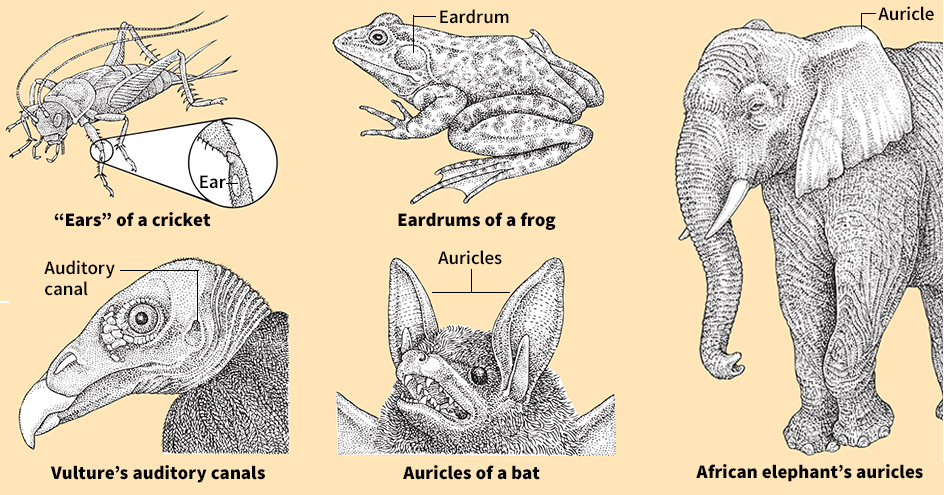
Only a few kinds of insects have true hearing organs. Their “ears” consist simply of thin membranes that vibrate when sound waves strike them. Among different insects, the “ears” are on the legs, sides, or other parts of the body. See Insect (Hearing).
Fish do not have any outer ears or eardrums. But some fish have a simple type of inner ear on each side of the head. These fish can hear sound waves that travel through the water. Sound vibrations are carried to the inner ear by a gas-filled sac called the air bladder. Some types of fish have a chain of ossicles that connect the air bladder to the inner ear. See Fish (Hearing) (Other senses).
Frogs and other amphibians have middle ears and inner ears. The middle ear of a frog consists of an eardrum and a small chamber with one bony ossicle. The eardrum of a frog is a large exposed disk behind the eye on each side of the head. See Frog (Senses).
Like amphibians, most reptiles have eardrums, middle ears, and inner ears. In some reptiles, the inner ears are highly developed. Most snakes lack eardrums, but they are not deaf, as many people think. Sounds are transmitted to the inner ears of snakes chiefly by the bones of the skull. See Reptile (Sense organs); Snake (Sense organs).
Birds have external auditory canals, middle ears, and inner ears. The cochlea of the bird inner ear is slightly curved but not coiled. The hearing ability of birds is similar to that of human beings. See Bird (Senses).
All mammals have coiled cochleas. In addition, mammals are the only animals that have auricles. In many mammals, these outer ear parts are movable and help channel sound waves into the auditory canal. African elephants have the largest auricles of any animal. Their auricles measure up to 4 feet (1.2 meters) wide. The auricles help the animals cool off in hot weather. Heat escapes from the elephant’s body partly through the skin of the auricles. Other animals, including certain kinds of rabbits and foxes, have unusually large auricles that serve the same purpose. See Mammal (Senses).
A few animals, including dolphins, some bats, and some whales, depend on hearing to navigate in the dark. They find their way about by a process called echolocation. The animals make sounds and listen for the echoes that are produced when objects reflect the sounds. From the echoes, they determine the distance to an object and the direction in which it lies. See Bat (Echolocation); Dolphin (The bodies of dolphins); Whale (Senses).
