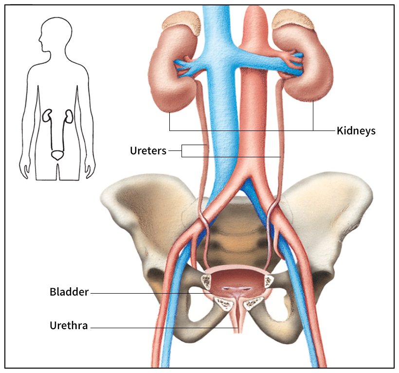
Bladder is the common name for the urinary bladder, a hollow muscular organ that stores urine before expelling it from the body. The emptying of the urinary bladder is voluntarily controlled in most human beings and many other mammals.

The bladder lies just behind the pubis, one of the bones of the pelvis. Urine drains continuously from the kidneys into the bladder through two tubes called ureters. It leaves the bladder through the urethra, a wider tube that leads out of the body. The place where the bladder and the urethra meet is called the neck of the bladder. A complex arrangement of muscles encircles the bladder neck. This ring, called the urethral sphincter, normally prevents urine from leaving the bladder.
The bladder can hold more than a pint (0.5 liter) of urine. As the bladder fills, its muscular wall relaxes and its lining stretches, allowing it to expand. The bladder begins to send signals to the brain that cause the desire to urinate. For urination to occur, the urethral sphincter must relax. The muscles of the bladder wall then contract, forcing urine out through the urethra.
The inability to control urination is called incontinence. In adults, incontinence may result from muscle weakness due to aging or from a variety of other causes. These include injury to the sphincter during surgery, damage to the bladder nerves, or a stroke affecting the brain’s regulation of urination.
Common diseases of the bladder include bladder inflammation, called cystitis, and cancer. Most cases of cystitis result from bacterial infection and can be cured with medication. If left untreated, the infection may spread to the kidneys. Cancerous tumors must be removed surgically. A physician uses an instrument called a cystoscope to look inside the bladder and to remove small tumors. A cystoscope is inserted through the urethra. Larger tumors may require removal of most or all of the bladder. The urine is rechanneled through an artificial opening in the abdomen and is collected in a plastic pouch worn by the patient.
