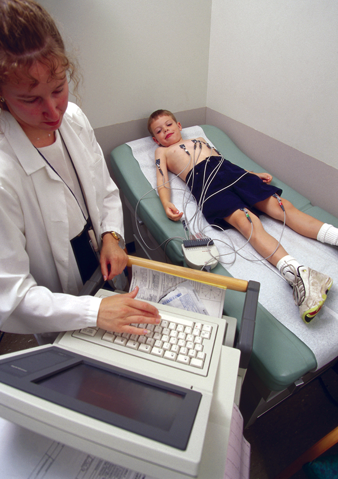
Electrocardiograph is an instrument used to diagnose heart disorders. Each time the heart beats, it produces electrical currents. These currents are responsible for the rate and pattern of contraction of the heart. An electrocardiograph picks up and records these currents. The electrocardiograph may be connected to a printer, which prints a record called an electrocardiogram, often abbreviated ECG or EKG. The electrocardiograph may also be connected to an oscilloscope, an instrument that displays the currents on a TV-type screen.

An electrocardiograph contains amplifying and recording equipment. Wires run from the machine to electrodes–strips of metal that conduct electricity. The electrodes are attached to the patient’s skin with a special jelly. Electrodes are placed on each arm and leg and at six points on the chest over the area of the heart.
The electrodes pick up the currents produced by the heartbeat and transmit them to an amplifier inside the electrocardiograph. The amplified currents then flow through a very fine wire coil that hangs suspended in a magnetic field. As these currents react with the magnetic field, they move the wire. In most electrocardiographs, a sensitive lever traces the wire’s motions on a moving paper chart, producing the electrocardiogram.
Each heartbeat produces a series of wavy lines on the ECG. A normal heartbeat makes a specific pattern of waves. Certain kinds of heart damage and disease change the pattern in recognizable ways.
Physicians use ECG’s to diagnose heart damage from such conditions as high blood pressure, rheumatic fever, and birth defects. An ECG also helps determine the location and the amount of injury caused by a heart attack, and follow-up ECG’s show how the heart is recovering. An ECG can also reveal irregularities in the heart’s rhythm, known as arrhythmias. In addition, physicians sometimes use an ECG to determine the effects of certain drugs on the heart.
An ECG is usually taken while the patient is lying down. This procedure is called a resting ECG. Sometimes, an ECG is taken while the patient is exercising or after the patient has received medication to stimulate the heart. This test, called a stress ECG, indicates whether the heart receives enough oxygen during vigorous activity. Doctors use a stress ECG to diagnose coronary artery disease. See Heart (Coronary artery disease) (Heart attack).
The electrocardiograph developed from the string galvanometer, invented in 1903 by the Dutch physiologist Willem Einthoven (see Einthoven, Willem; Galvanometer). The electrocardiograph was first used in the United States in 1909.
See also Stress test.
