Magnetic resonance imaging (MRI) is a technique used in medicine and research for producing images of tissues inside the body. MRI is an important diagnostic tool because it enables physicians to identify abnormal tissue without opening the body through surgery. MRI does not expose the patient to radiation, unlike tests that use X rays. In medicine, MRI is used for diagnosing diseases, disorders, and injuries, and helps physicians determine treatment. An MRI examination or scan is supervised by a radiologist—a physician trained in using images for medical diagnosis. MRI is used to view the structures of the head and spine and to detect tumors and other abnormalities in the chest, abdomen, joints, and other parts of the body.
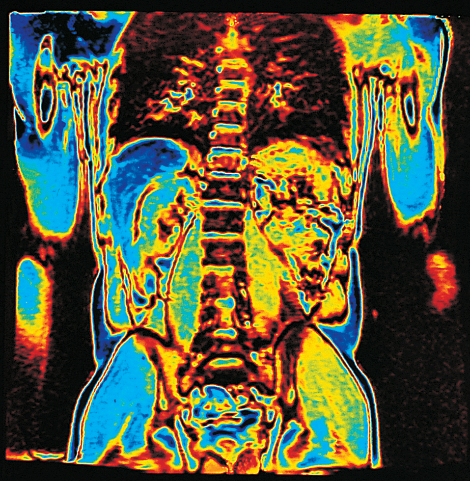
An MRI system is made up of one or more large magnets as well as computers and devices for transmitting and receiving radio waves. During the MRI scan, a magnetic field is applied to the person’s body. The magnetic field causes nuclei in certain atoms in the body, mainly atoms of water, to line up. Radio waves are then directed at the nuclei. If the frequency of the radio waves equals that of the atom, a resonance condition results. This condition enables the nuclei to absorb the energy of the radio waves. When the radio-wave stimulation stops, the nuclei return to their original state and emit energy in the form of weak radio signals. The strength and length of these signals—and therefore the kind of image produced—depends on the properties of the tissue. For example, the signal generated in portions of the body containing fluid is different from the signal generated by portions composed of fat. Computers translate the signals into detailed images. In some imaging procedures, patients may receive an injection of a contrast agent, a chemical that enhances the resulting image.
MRI can measure the diffusion (spread) of water molecules in the body. This information is used to construct images that indicate the connections and directions of nerve fibers in the brain. With these images, scientists can study the structure of the healthy brain and the diseases that damage nerve tissue, such as multiple sclerosis. They can also identify areas of restricted diffusion in brain tissue, which occurs in a type of stroke called ischemic stroke, where a blood vessel in the brain becomes blocked.
A more specialized form of MRI called functional MRI (fMRI) can provide information about brain function as opposed to conventional MRI techniques, which only image brain anatomy. Functional MRI is used to detect and view areas of increased blood flow and blood volume in the brain. Scientists know that blood flow increases in regions of the brain where brain cells are active. Increases in blood flow in a particular region of the brain observed with fMRI suggest activity there. Research scientists use fMRI to reveal which regions of the brain are active during particular mental operations.
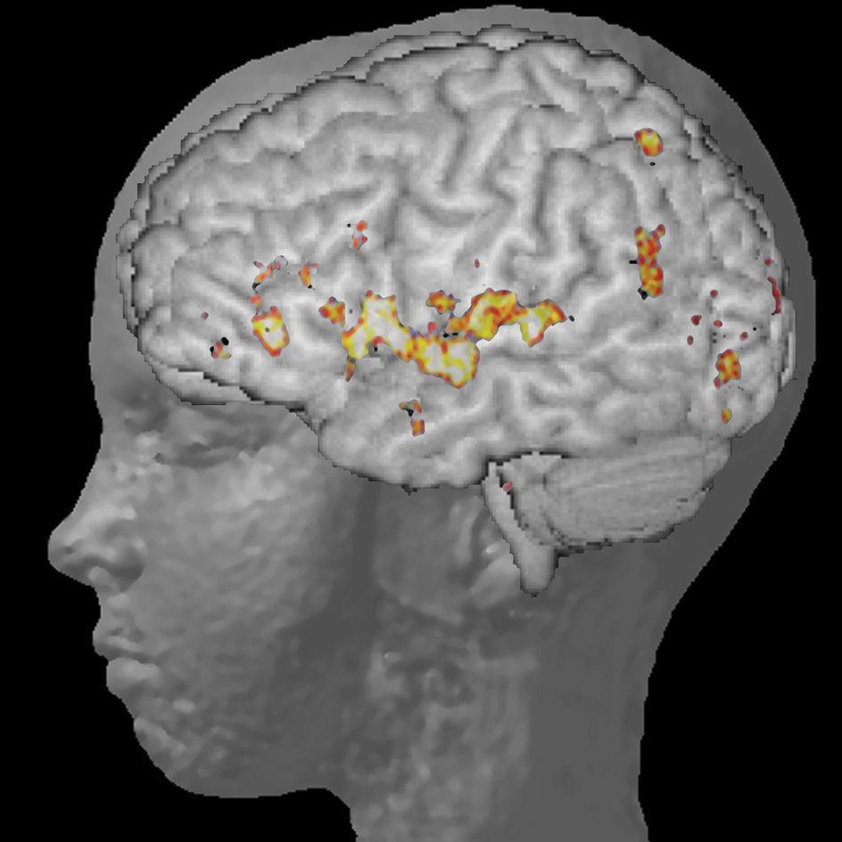
Unlike other medical imaging techniques, such as positron emission tomography (PET), fMRI does not require the injection of radioactive materials to produce images. This makes fMRI more suitable for repeated scanning and for children. Functional MRI also typically produces images of a higher resolution than PET does.
MRI devices have a strong magnetic field, the strength of which is measured in units called gauss or tesla. One tesla equals 10,000 gauss. Earth’s magnetic field at its surface is about 1/2 gauss. Fields of magnets used in industry may measure more than 2 tesla (20,000 gauss). Hospitals use MRI systems of 0.5 to 3 tesla (5,000 to 30,000 gauss), and medical scientists use systems of about 1.5 to 9.0 tesla (15,000 to 90,000 gauss) for research.
Most MRI devices are so large and have such a strong magnetic field that they need to be housed in special facilities, such as clinics, medical centers, and research institutes. However, smaller, less powerful, and portable MRI units can be used for some imaging procedures. Nevertheless, the strength of the magnetic field is so great, even in the smaller units, that MRI cannot be used on people with metal implants, such as pacemakers or certain artificial joints.
MRI technology is used outside medicine in various areas of science and research, particularly chemistry. A specialized form of this technology called nuclear magnetic resonance (NMR) spectroscopy helps scientists determine the chemical makeup and molecular structure of substances. NMR spectroscopy is often used in the pharmaceutical industry to analyze new chemical compounds.