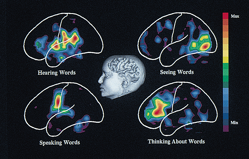
Positron emission tomography, << tuh MOG ruh fee >> (PET), is a technique used to produce images of the chemical activity of the brain and other body tissues. PET enables scientists to observe chemical changes in specific regions of a person’s brain while the person performs various tasks, such as listening, thinking, or moving an arm or leg. Scientists use PET to compare the brain processes of healthy people and people with diseases of the brain. Research is being done to see if it is possible to use these comparisons to identify abnormalities that underlie various brain disorders. These disorders include such mental illnesses as bipolar disorder and schizophrenia, as well as such conditions as Alzheimer’s disease, cerebral palsy, epilepsy, and stroke. PET also helps doctors diagnose certain other disorders, including heart disease and cancer.

In a PET scan of the brain, the patient’s head is positioned inside a ring of cameralike sensors. These sensors can detect gamma rays (short-wave electromagnetic radiation) from many angles. A solution containing a harmless amount of a radioactive element bound to glucose is injected into a vein. This radioactive labeled glucose mixes with the glucose in blood and soon enters the brain.
The radioactive substance gives off positrons, particles identical to electrons but carrying an opposite electric charge. The positrons collide with electrons present in brain tissue and gamma rays are emitted (given off). The sensors record the points where these rays emerge. A computer then assembles these points into a three-dimensional representation of the emitting regions. This representation is displayed on a video screen as cross-sectional “slices” through the brain.
Colors in a PET image show the rate at which specific brain structures consume the glucose. The rate of glucose consumption indicates how active these structures are during a particular task. For example, if the person having the PET scan looks at an object, the brain region that receives and interprets visual signals will appear red on the screen. Red indicates the highest rate of activity. Other colors that appear in PET images include orange—the next highest rate of activity—yellow, green, and blue, the lowest rate.
