Teeth are hard, bonelike structures in the upper and lower jaws of human beings and many kinds of animals. They are the hardest parts of the body.
People use their teeth chiefly to chew food. Chewing is the first step in the process of digestion. Digestion begins as the teeth chop and grind chunks of food into smaller pieces. As the teeth chew the food, it is mixed with saliva, a liquid produced in the mouth. The food becomes a moist pulp, which is easy to swallow. The food is further broken down in the stomach and the small intestine, where it is absorbed by the blood. The blood carries the digested food to all parts of the body. Without teeth, people could not eat foods that must be chewed. They could only swallow soft foods and liquids.
Teeth also play an important part in speech. The teeth and tongue are used together to form many sounds that make up words. To produce the th sound, for example, the tip of the tongue is placed against the upper front teeth. A person who lacks these teeth may be unable to make the sound. Teeth also help support the muscles around the mouth and so contribute to a person’s appearance. People who have lost their teeth lack this support. Unless they wear artificial teeth, they may have deep, saggy lines around the mouth.
Like human beings, most animals use their teeth to chew food. They also use their teeth to obtain food. Many animals that eat plants tear off the leaves or stalks of the plants with their teeth. Most meat-eating animals use their teeth to seize and kill prey.
Kinds of teeth
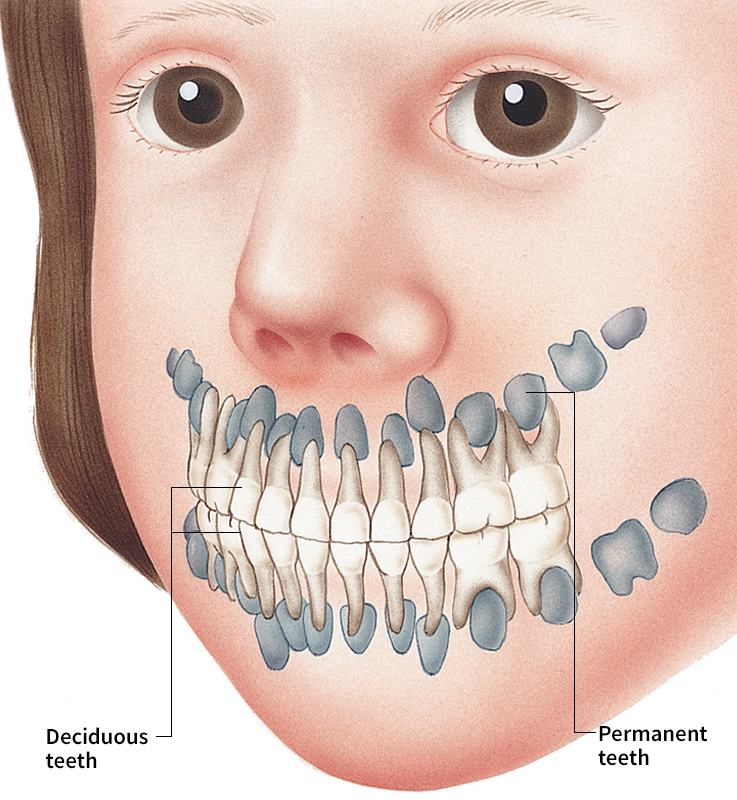
Human beings grow two sets of teeth: (1) deciduous teeth and (2) permanent teeth. The individual deciduous teeth appear and fall out gradually early in life. They are replaced, one by one, by the permanent teeth. See the table “Ages at which teeth appear” for the times deciduous and permanent teeth generally appear.
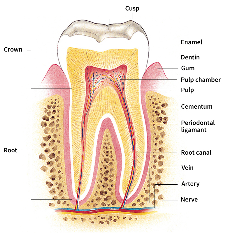
Deciduous and permanent teeth have the same basic structure. Each tooth has a crown and one or more roots. The crown is the part of the tooth that can be seen in the mouth. Bone and gums cover the root or roots. The roots hold the tooth in a socket in the jawbone.
Deciduous teeth
are also called baby teeth, milk teeth, or primary teeth. They start to form about 71/2 months before a baby is born. They begin as oval or round swellings called buds, which gradually develop into teeth. When a baby is born, parts of all the deciduous teeth are present deep within the jaws. As the teeth grow, they push through the gums. This process is called eruption or teething. Babies begin to teethe at about 6 to 9 months of age. Most children have all their deciduous teeth by about 2 years of age.
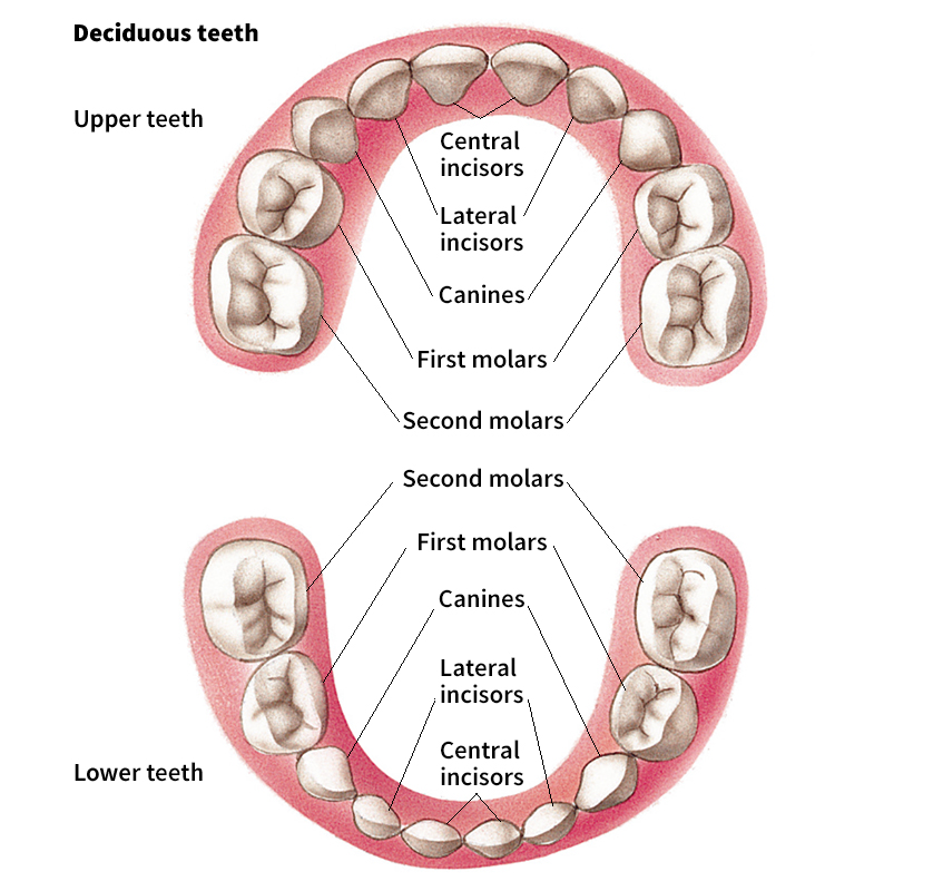
There are 20 deciduous teeth, 10 of them in each jaw. They consist of three kinds of teeth: (1) incisors, (2) canines, and (3) molars. Each jaw has 4 incisors, 2 canines, and 4 molars. The incisors and canines are used to bite into food, and the molars are used to grind food. The positions of all the deciduous teeth in the mouth are shown in the illustration “Kinds of teeth.”
The deciduous teeth help the permanent teeth erupt in their normal positions. Most of the permanent teeth form near the roots of the deciduous teeth. When a child is about 3 years old, the roots of various deciduous teeth begin to dissolve slowly. By the time a permanent tooth is ready to erupt, the root of the deciduous tooth has completely dissolved. The crown of the tooth then becomes loose and falls out. 
Permanent teeth,
like deciduous teeth, begin to develop before birth. But most of their growth occurs after birth. The permanent teeth begin to erupt after the deciduous teeth start to fall out. The first permanent teeth appear when a child is about 6 or 7 years old. Between the ages of 6 and 12, a child has some permanent and some deciduous teeth in the mouth. The last permanent teeth erupt when a person is 17 to 21 years old.
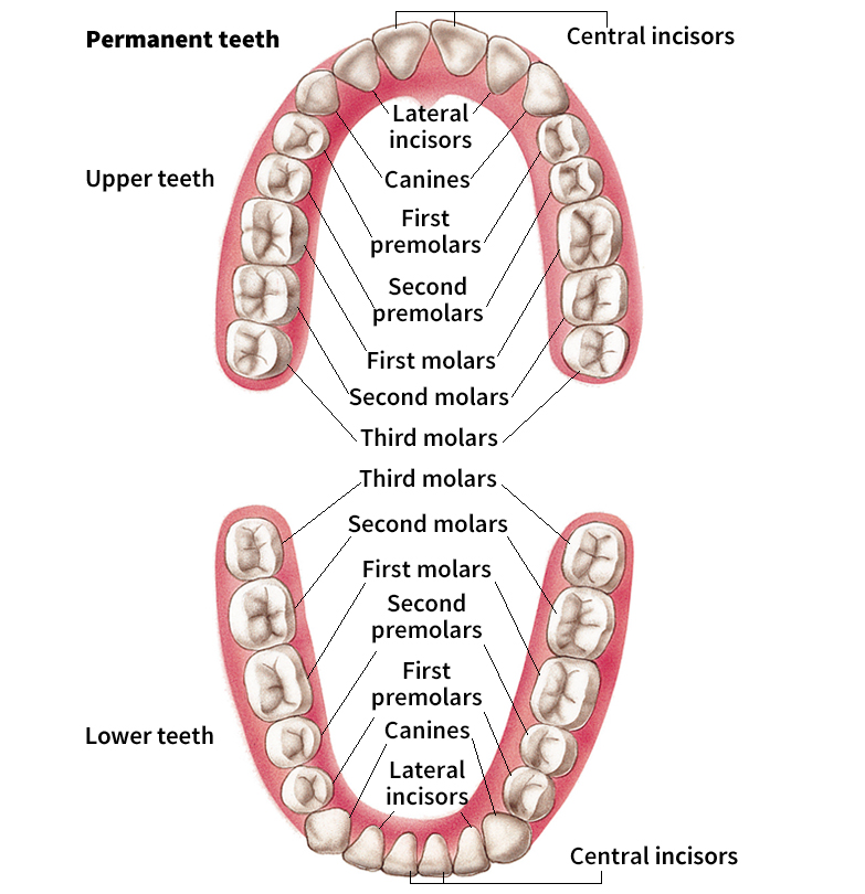
There are 32 permanent teeth, 16 in each jaw. They are larger than the deciduous teeth and consist of four kinds of teeth. The four kinds are (1) incisors, (2) canines, (3) premolars, and (4) molars. Each jaw has 4 incisors, 2 canines, 4 premolars, and 6 molars. The following discussion describes the four kinds of permanent teeth. Their positions in the mouth are shown in the illustration “Kinds of teeth.”
Incisors are the chief biting teeth. They have a sharp, straight cutting edge. In most cases, incisors have one root. The central incisors of the lower jaw are the smallest permanent teeth.
Canines are used with the incisors to bite into food. They are also used to tear off pieces of food. The name for these teeth comes from canis, the Latin word for dog, because they resemble some of a dog’s teeth. They have a sharp, pointed edge and one root. Canines are also called cuspids or dogteeth. The upper canines are sometimes known as eyeteeth.
Premolars are used to crush and grind food. They have a broad, lumpy top instead of a sharp biting edge. The small surface lumps are called cusps. The cusps enable the teeth to mash pieces of food.
Premolars are sometimes called bicuspids because, in most cases, they have two cusps. The prefix bi means two. The first upper premolars normally have two roots. The other premolars have one root. The premolars erupt in the place of the deciduous molars.
Molars, like premolars, are used to grind food. They are shaped much like premolars but are larger. The various molars normally have three to five cusps and two or three roots.
The permanent molars do not form beneath any of the deciduous teeth. They develop as the jaws grow, which makes space for them. Some adults lack one or more of the third molars, which are commonly called wisdom teeth. In many cases, the jaws do not grow large enough to provide space for the wisdom teeth. As a result, the wisdom teeth may become impacted—that is, wedged between the jawbone and another tooth. The wisdom teeth must then be removed.
Parts of a tooth
A tooth consists of four kinds of tissues. They are (1) pulp, (2) dentin, (3) enamel, and (4) cementum. Connective tissue surrounds the root of the tooth. This tissue, called the periodontal ligament, holds the root in the socket in the jaw.
Pulp
is the innermost layer of a tooth. It consists of connective tissue, blood vessels, and nerves. The blood vessels nourish the tooth. The nerves transmit sensations of pain to the brain.
The pulp has two parts, the pulp chamber and the root canal. The pulp chamber lies in the crown of the tooth. The root canal lies in the root of the tooth. Blood vessels and nerves enter the root canal through a small hole at the tip of the root. They extend through the root canal and into the pulp chamber.
Dentin
is a hard, yellow substance that surrounds the pulp. It makes up most of a tooth. Dentin is harder than bone. It consists mainly of mineral salts and water but also has some living cells.
Enamel
overlies the dentin in the crown of the tooth. It forms the outermost covering of the crown. Enamel is the hardest tissue in the body. It enables a tooth to withstand the pressure placed on it during chewing. Enamel consists of mineral salts and a small amount of water. Enamel is white but transparent. The yellow color of the dentin shows through the enamel, and so most teeth appear slightly yellowish.
As a person grows older, small amounts of enamel begin to wear away. This process, called attrition, results from the use of the teeth over a long period. As the enamel wears away, the dentin becomes exposed.
Cementum
overlies the dentin in the root of the tooth. In most cases, the cementum and enamel meet where the root ends and the crown begins. As the surface of the tooth wears away, the tooth grows farther out of its socket, exposing the root. These areas may then become more sensitive to hot and cold liquids. Cementum is about as hard as bone. Like dentin and enamel, it consists mainly of mineral salts and water.
Periodontal ligament
consists of small fibers. These fibers extend through the cementum and into the bony socket, which is called the alveolus. Besides anchoring the tooth in the alveolus, the periodontal ligament serves as a shock absorber during chewing.
Care of the teeth and gums
Most cases of tooth decay and gum disease could be prevented if people took proper care of their teeth and gums. Proper care requires (1) a good diet, (2) cleaning the teeth after eating, and (3) dental checkups.
A good diet.
Dentists advise people to eat well-balanced meals. Such meals include a variety of foods and provide the nutrients (nourishing substances) needed by the teeth and gums. Nutrition experts divide foods into groups to help people plan well-balanced meals. The article Nutrition describes the Food Pyramid, developed by the U. S. Department of Agriculture (USDA). The pyramid shows the recommended number of daily servings of the major food groups in a healthful diet.
Dentists also urge people to eat fewer sugary foods because these foods contribute to tooth decay. Bacteria in the mouth digest sugar and produce an acid as a result. The acid dissolves tooth enamel, forming a cavity.
Foods that have a large amount of sugar include candies, pastries, most breakfast cereals, and sweetened canned fruits. Many people eat sugary foods as snacks. In place of sugary foods, dentists advise people to snack on such foods as fresh fruits and vegetables, cheeses, and nuts. They also recommend that people drink skim milk or unsweetened fruit and vegetable juices instead of soft drinks and other sugar-sweetened beverages.
Dentists further recommend that children drink water with chemical compounds called fluorides. Fluorides are absorbed by the enamel as the teeth grow. They help the teeth resist acid that forms cavities. Some communities have a water supply that naturally contains fluorides. Many other communities add fluorides to the water supply. But some people oppose fluoridation (the addition of fluorides to water supplies). For information on the arguments, see Fluoridation.
Fluorides may be applied directly to a child’s teeth during a dental checkup. In some cases, dentists prescribe a fluoride substance that children can apply at home. Most dentists also advise children to brush their teeth with a toothpaste that contains fluorides.
Cleaning the teeth.
Dentists advise people to clean their teeth by brushing after every meal and by using dental floss once a day. Dental floss is a thin thread that comes in a roll. It is used to clean the areas between teeth and under the gum line. Brushing and flossing remove trapped food particles and plaque from the teeth. Plaque is a sticky film that consists of saliva, food particles, and bacteria. The bacteria digest certain foods, particularly sugars, and form an enamel-dissolving acid.
To brush the teeth, you should use a small, soft toothbrush and a toothpaste that contains fluorides. There are several methods of brushing. You should use the one recommended by your dentist. One commonly recommended method is to place the brush against the teeth at a slight angle, with the bristles pointed toward the gums. Brush the upper teeth with a downward, sweeping motion. Brush the lower teeth with an upward, sweeping motion. Clean both the outside and the inside surfaces of the teeth in this way. Use a scrubbing motion to clean the biting surfaces of the premolars and molars. Lastly, brush the tongue to remove food particles and bacteria, which contribute to bad mouth odors. Then rinse the mouth thoroughly with water or mouthwash. 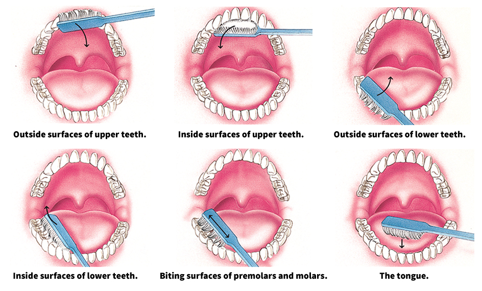
To floss the teeth, cut a piece of floss about 18 inches (46 centimeters) long from the roll. Wrap one end of the floss around each middle finger. Using the index fingers and thumbs, gently guide the floss between two teeth. Then pull the floss up and down, cleaning the sides of both teeth and the areas around the gum line. Repeat this procedure on all the teeth.
Some people use disclosing tablets to see if any areas of the teeth remain unclean after brushing and flossing. Disclosing tablets contain a red or purple dye. When you chew a tablet, the dye sticks to unclean areas of the teeth. You can then rebrush and refloss these spots.
Dental checkups.
Dentists advise people to have a dental checkup at least once a year. Children should start going to a dentist after all their deciduous teeth have erupted. Dentists can recognize and treat diseases of the teeth and gums at an early stage, before the diseases cause serious damage. Dentists also provide services that help prevent diseases of the teeth and gums. Many dentists employ a licensed dental hygienist to help them in their work.
During a checkup, the dentist looks at the teeth, gums, and other tissues inside the mouth for signs of diseases. The dentist—or the dental hygienist—also may X-ray the teeth. X rays can show the location of dental decay that cannot be seen. They also show any abnormal conditions of the jawbones and other tissues that support the teeth. Based on the examination, the dentist may decide to fill cavities or plan other treatment. The dentist or hygienist then cleans the teeth to remove plaque and calculus, a hard, yellowish substance formed by the buildup of plaque. Calculus is also called tartar. After the teeth have been cleaned, a fluoride substance is applied to help the teeth resist decay. Generally, only children and teenagers receive applications of fluorides. Lastly, the dentist or hygienist may instruct the patient on how to brush and floss the teeth properly. 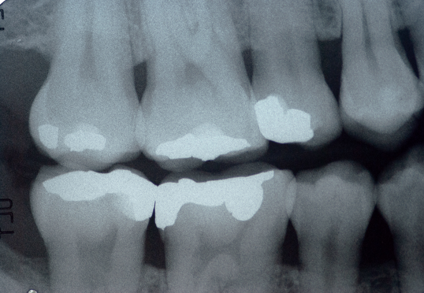
Diseases and defects of the teeth
Dental decay, also called caries, is the most common disease of the teeth. Most people under the age of 35 who lose their teeth do so because of dental decay. A defect in the position of the teeth, called malocclusion, is also a common problem among young people. Diseases of the gums and alveolus, called periodontal diseases, are the chief dental problem of people over the age of 35. A less common but very severe disease is oral cancer, which kills about 8,000 people in the United States each year. The following discussion describes the causes and treatment of (1) dental decay, (2) malocclusion, (3) periodontal diseases, and (4) oral cancer.
Dental decay
is a complex process that involves plaque, bacteria, and food. Saliva produces an invisible film on the teeth. Bacteria and food particles stick to this film, forming plaque. The bacteria digest the carbohydrates (sugars and starches) in food and produce an acid. The acid dissolves enamel, causing a cavity. If the cavity is not treated, the decay will progress through the enamel and into the dentin. When the decay reaches the pulp, a toothache results. Loading the player...
Tooth decay
The occlusal (biting) surfaces of the premolars and molars tend to decay easily because they have many small pits, which trap food. This type of decay may be prevented in deep pits by applying a surface sealant. The sealant is a plasticlike material and is bonded to the occlusal surface.
Dentists have several methods of treating dental decay, depending on the severity. The most common methods include (1) filling a cavity, (2) performing root canal therapy, (3) crowning a tooth, and (4) removing and replacing teeth. Before beginning any of these procedures, the dentist usually injects an anesthetic (painkilling drug) into the gums near the nerves of the tooth. In some procedures, electronic anesthesia may be used. This technique sends gentle electronic impulses to the brain through electrodes (conductors) positioned in the mouth. These impulses block pain signals. The patient uses a handheld remote control unit to increase or decrease the amount of anesthesia.
Filling a cavity.
To fill a cavity, the dentist first removes the decayed and soft parts of the tooth, using small hand instruments, a laser, or an air-powered or electric drill. The dentist then makes tiny undercuts or ledges in the hole. These undercuts help hold the filling, which is not adhesive. The filling is packed into the hole and allowed to harden slightly. The dentist then carves the filling to restore the original shape of the tooth. In most cases, dentists fill cavities with silver amalgam or gold. Silver amalgam consists of a mixture of silver and mercury and a small amount of copper and tin. 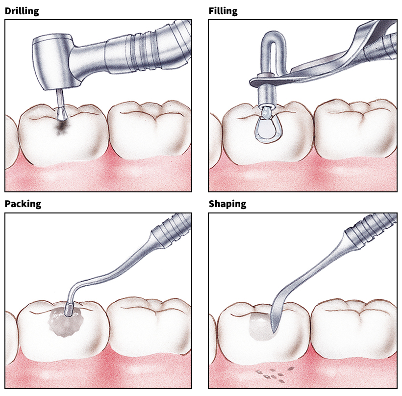
Another method of filling cavities uses a plasticlike resin that is shaded to match the tooth color. This method is particularly useful in repairing decay or breaks in the front teeth. After any decay has been removed, a small amount of dilute acid is applied to the area to be filled. The acid etches or grooves the surrounding enamel surface and is left in place for about one minute. The acid is then rinsed off with water and the area is dried. The resin is then applied to the etched area and formed into the shape of the cavity or break. After hardening, the resin is shaped and polished with a drill.
Performing root canal therapy.
Root canal therapy is the removal of the pulp of a tooth. It is performed if the pulp has become infected. When decay extends into the pulp, a small sac of pus, called an abscess, may form. An abscess can be extremely painful. If it is not treated, infection may spread to other parts of the body.
To perform root canal therapy, the dentist first anesthetizes the area and then drills a hole into the crown of the tooth. The dentist uses small files to reach through the hole and clean out the pulp. After removing the pulp, the dentist fills the empty space, usually with a rubberlike substance called gutta-percha. Sometimes, the hole in the crown of the tooth is then filled. But in the majority of cases, the tooth must be fitted with an artificial crown.
Crowning a tooth.
Crowns are toothlike caps that may be made of metal, porcelain, or plastic. They are used when the natural crown is so badly damaged that it does not have enough healthy tissue to hold a filling. 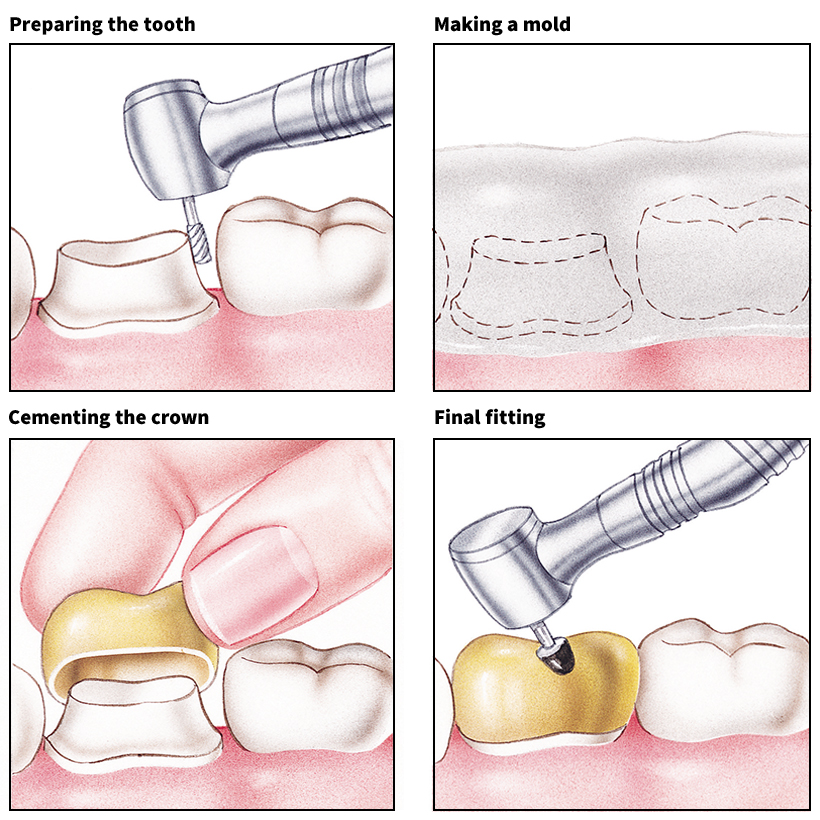
To crown a tooth, the dentist first anesthetizes the area and then prepares the natural crown by grinding it down slightly. Next, the dentist covers the prepared tooth and the teeth next to it with a jellylike material. After this material hardens, it is removed from the patient’s mouth and serves as an impression (mold). The dentist also makes an impression of the teeth in the opposite jaw that press against the prepared tooth and the teeth next to it. The impressions are used to make a plaster reproduction of the prepared tooth and other teeth. Dental technicians then produce a crown, using the plaster reproduction as a model. They must make sure that the crown not only fits the prepared tooth but also fits in place with the other teeth.
Meanwhile, a temporary crown is placed on the tooth. When the permanent crown is ready, the dentist removes the temporary crown and cements the permanent one onto the tooth.
Crowns can also be made by a computerized method that eliminates the need for impressions and plaster models. This method is called computer-aided design and computer-aided manufacturing (CAD/CAM). The dentist uses an optical probe system to record electronic images of the tooth. This information is entered into a computer, which designs an electronic model of the tooth. The design is then transferred through the computer to the head of a cutting tool, which prepares the crown to exactly fit the tooth.
If a tooth is only slightly damaged, a dentist may cover the tooth with thin porcelain sheets called veneers. The veneers are made to match the shape and color of a tooth and are bonded to the tooth using a cementlike material. They are used primarily to improve the appearance of front teeth.
Removing and replacing teeth.
In severe cases of dental decay, a dentist may remove one or more teeth and replace them with artificial ones. But artificial teeth do not function as well as natural teeth. Dentists therefore remove teeth only if no other method of treatment is considered possible. To remove a tooth, a dentist first anesthetizes the area. The dentist uses an instrument that resembles a pliers to grip the crown of the tooth and loosen the root from the socket. Both the crown and the root are then removed. After the gums heal, the patient can be fitted with an artificial tooth.
Artificial teeth are made from impressions taken of the patient’s mouth. In most cases, the teeth are made of plastic. The most common types of artificial teeth are bridges, partial dentures, and full dentures. Bridges are permanently fixed in the mouth, but partial dentures and full dentures are removable. Bridges are used when only a few teeth are missing. They consist of one or more artificial teeth with a metal or porcelain crown on each side. The crowns fit over the adjoining natural teeth, which must be prepared to hold the crowns. Partial dentures are also used to replace only a few missing teeth. A partial denture has metal clasps that hook around nearby teeth and hold the denture in place. Full dentures are used when all the teeth of one or both jaws are missing. In a full denture, the artificial teeth are attached to a plastic base that fits over the ridge left after the teeth have been removed. In the upper jaw, the plastic base also covers the roof of the mouth.
Another method of replacing one or more missing teeth involves the use of dental implants. In the most common implant procedure, metal or ceramiclike cylinders, called fixtures or root forms, are surgically implanted in the area of the jaw where the teeth are missing. After the area has healed, artificial teeth are permanently attached to the root forms. Implants cannot be used if the jaw area is severely damaged.
Malocclusion
is the failure of the teeth in the upper and lower jaws to meet properly when a person bites. Normally, the upper front teeth should slightly overlap the lower front teeth. There are three main types of malocclusions, overbite, underbite, and crowding. In overbite, the upper front teeth stick out too far over the lower front teeth. This defect is commonly called buck teeth. In underbite, the lower front teeth extend in front of the upper ones. Many people have the correct occlusion (bite), but their teeth are crowded. Crowding is the most common malocclusion. 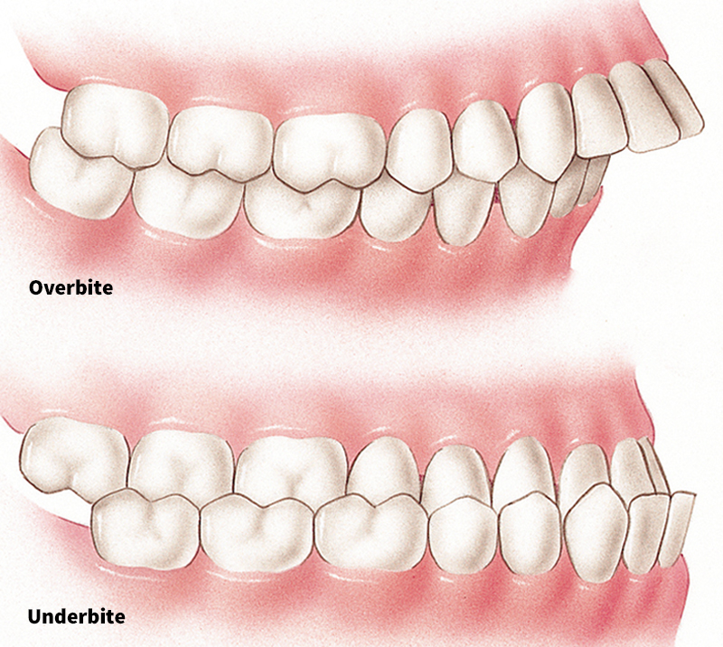
Malocclusion has various causes. In some cases, a deciduous tooth falls out before a permanent tooth is ready to erupt. The nearby teeth then gradually move into the open space and prevent the permanent tooth from erupting in the correct position. In other cases, the permanent teeth are too large for the jaw and crowd one another. The edges of some teeth may then overlap, or one tooth may grow above another. In still other cases, the jaws do not grow properly.
Malocclusion prevents the teeth from functioning normally when a person chews food. It also may affect the way a person speaks. In addition, malocclusion contributes to the development of dental decay and periodontal diseases, partly because irregularly positioned teeth are hard to clean.
Most cases of malocclusion can be corrected with braces. Braces consist of metal or clear ceramic brackets that are bonded on the front surface of each tooth and connected by wires. The wires are tightened periodically to force the teeth to move into the correct position. But the teeth must be moved slowly, and so the treatment may take a year or more. In some cases, one or more teeth must be removed to allow enough space for the others to move into a normal position. 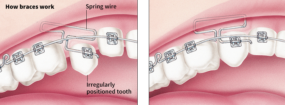
Periodontal diseases
are caused chiefly by the build-up of plaque and calculus between the gums and teeth. The plaque and calculus irritate the gums, causing them to become inflamed. In time, the jawbones may become infected. The best way to prevent plaque from building up under the gum line is by flossing daily. The gums can also become irritated by habitually breathing through the mouth, smoking or chewing tobacco, brushing improperly, or wearing ill-fitting dentures. Irregularly positioned teeth can also irritate the gums. 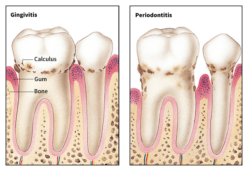
There are three main kinds of periodontal diseases. They are (1) gingivitis, (2) periodontitis, and (3) Vincent’s infection.
Gingivitis
is an inflammation of the gingivae (gums). The gingivae become red and swollen and bleed easily when brushed or prodded. Dentists treat gingivitis by cleaning the teeth and gums to remove plaque and calculus. They also instruct patients on how to brush and floss the teeth and on how to massage the gums. If gingivitis is not treated, it can lead to periodontitis.
Periodontitis,
also called pyorrhea, is a severe infection of the gingivae, alveolus, and other tissues that support the teeth. The infection gradually destroys the bony walls of the sockets, and the teeth become loose. Periodontitis is difficult to cure, but it can be effectively controlled. Treatment involves surgical or nonsurgical removal of the damaged tissues and repair of the remaining healthy tissues. Some dentists use the heat of a beam from a laser to remove infected tissue. The laser process may quicken healing because it does little damage to the surrounding healthy area. Sometimes, loose teeth can be splinted (attached) to nearby teeth that are still firm. But in many cases, the loose teeth must be removed and replaced by artificial ones.
Vincent’s infection, also called trench mouth, is a painful infection of the gingivae. The gums become red and swollen and bleed easily. The mouth has an extremely bad odor, and the victim may develop a fever. To treat Vincent’s infection, a dentist cleans the teeth and gums thoroughly and instructs the patient on mouth care. In most cases, the dentist also prescribes antibiotics to combat the infection.
Oral cancer
is a disease that destroys the tissues of the mouth and may spread to other parts of the body. Scientists do not know for certain what causes oral cancer. But many factors can contribute to its development. For example, people who smoke or chew tobacco or drink excessive amounts of alcoholic beverages increase the risk of developing oral cancer.
Oral cancer may be painless and unnoticeable in its early stages. The first symptom may be a small sore in the mouth that does not heal. To test for cancer, a dentist removes some tissue from the sore. The tissue is examined under a microscope to determine if it is cancerous. Oral cancer may be treated with drugs, radiation, or surgery.
Teeth of animals
Many kinds of animals have teeth. However, birds, toads, turtles, and some types of insects and whales do not have teeth.
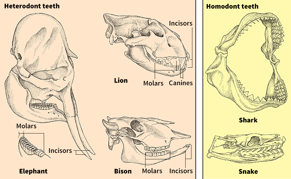
Cats, dogs, and most other mammals have heterodont teeth—that is, they have at least two types of teeth, which have different uses. For example, they may have incisors for biting into food and molars for crushing or grinding food.
The teeth of various kinds of mammals differ in shape and size, depending chiefly on what the animals eat. For example, plant-eating mammals, such as elephants, giraffes, and sheep, have unusually broad, flat molars. They use the molars to chew and mash plants. Meat-eating mammals, such as lions, tigers, and wolves, have long, pointed canines. They use the canines to rip and tear the bodies of their prey.
Some mammals have teeth that grow continuously. The tusks of elephants are actually incisors that have become very long. The tusks have an open pulp, which enables them to keep growing. Beavers, rats, and other rodents also have teeth that grow continuously. But most of the growth is worn down by continual use of the teeth, and so the teeth of these animals do not lengthen greatly.
Unlike most mammals, many fish and most reptiles have homodont teeth—that is, all their teeth are about the same size and shape and have only one use. In general, animals that have homodont teeth use their teeth to catch prey. Fish and reptiles lose and replace their teeth continuously.
Snakes have teeth that curve back toward the throat. Snakes swallow their prey whole and use their teeth to pull the prey back into the throat. In poisonous snakes, certain teeth have a canal or a groove, through which poison can be ejected. The poison comes from glands in the roof of the mouth.