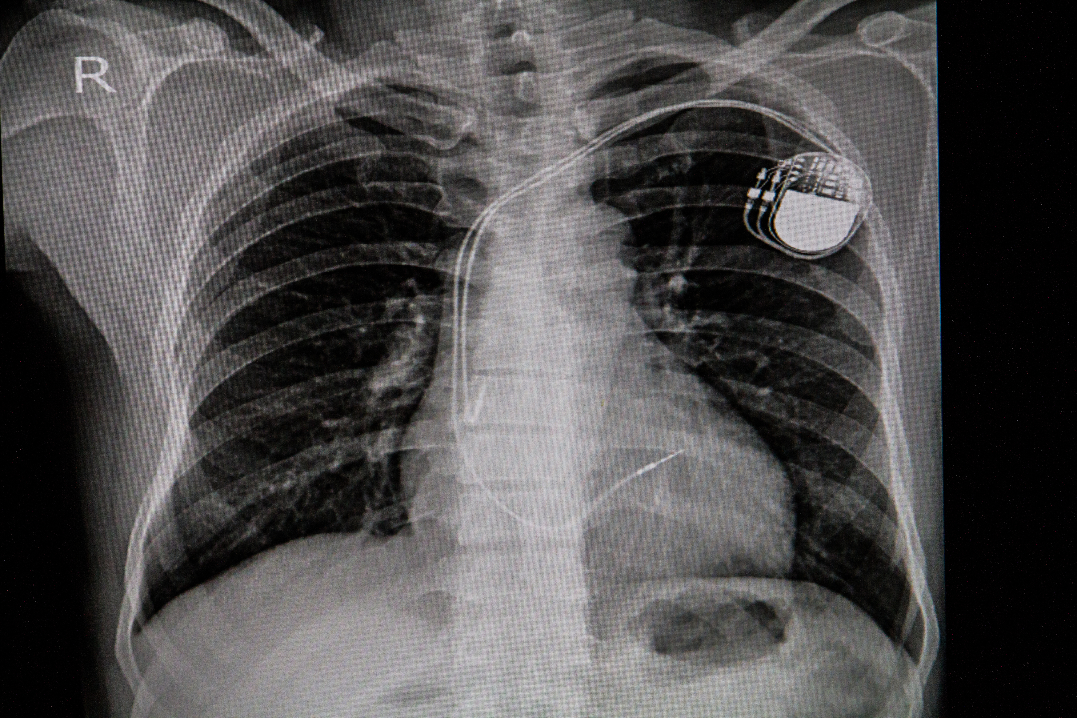
Pacemaker is a medical device used to regulate heartbeat. It is placed under the skin of the chest. A pacemaker is the size of a large coin. A person may need a pacemaker to control an arrhythmia (abnormal heart rhythm). Pacemakers monitor heart rate and correct heartbeat accordingly. For instance, if the heart rate becomes too slow, the pacemaker will send electrical signals that cause the heart to beat more rapidly.

An artificial pacemaker mimics the body’s natural pacemaker—the sinoatrial node. The sinoatrial node is a collection of cells that generate electrical signals to initiate heartbeat. It is located in the right atrium (upper heart chamber).
A pacemaker consists of three parts: (1) the computer; (2) the pulse generator; and (3) the leads, also called electrodes . The computer analyzes heart rate and determines when to send a corrective signal. The pulse generator includes a battery and electric circuits that create electrical pulses. Both the computer and the pulse generator are housed within the body of the pacemaker. The leads are wires that connect the pacemaker to the chambers of the heart, delivering the electrical pulses. A pacemaker has one to three leads.
There are three types of pacemakers: (1) single chamber, (2) dual chamber, and (3) biventricular. A single-chamber pacemaker delivers electrical signals from the pulse generator to the right ventricle (lower heart chamber). A dual-chamber pacemaker delivers electrical signals from the pulse generator to the right ventricle and the right atrium. A biventricular pacemaker delivers signals from the pulse generator to the right and left ventricles. Only people with extreme heart failure receive biventricular pacemakers.
Before receiving a pacemaker, a patient undergoes such tests as an electrocardiogram and a stress test to determine the nature of the arrhythmia. The surgery to implant a pacemaker takes several hours. The patient is given local anesthesia (pain-numbing medication) but remains awake during surgery. The surgeon inserts the leads into a major vein under or near the collarbone, then guides them to the heart. One end of each lead is secured to the heart. The other end is attached to the body of the pacemaker, which is placed under the skin beneath the collarbone.
Doctors and patients can monitor pacemakers remotely using wireless technology. They can receive information about heart rate and rhythm, pacemaker function, and battery life. Once implanted, the battery may last 5 to 15 years. If the battery runs low, doctors may replace the body of the pacemaker, connecting the new unit to the existing leads.
Electrical activity in the heart was discovered during the 1800’s, but medical devices to control heart rate were not produced until the 1900’s. The American physiologist Albert Hyman built the first pacemaker in 1932. A hand-cranked motor powered the device. After World War II (1939-1945), the American cardiologist (heart doctor) Paul Zoll and the American engineer Earl Bakken made smaller pacemakers, some of which could be worn like a necklace. In 1958 in Sweden, the first pacemaker was implanted in a patient, Arne Larsson. In the 1960’s, the British surgeon Leon Abrams and the British engineer Ray Lightwood created a pacemaker that became widely available to the public. Before the 1970’s, pacemakers had short-lived, unreliable batteries. This problem was solved in 1970, when the American engineer Wilson Greatbatch developed a lithium battery with a longer life.
